A History of Glasses
An interesting review of the history of glasses throughout time.
A video description

An interesting review of the history of glasses throughout time.
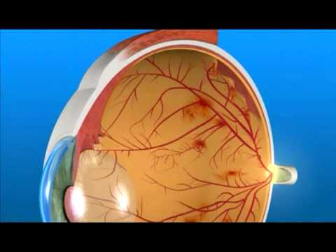
A patient’s guide to diabetic retinopathy.
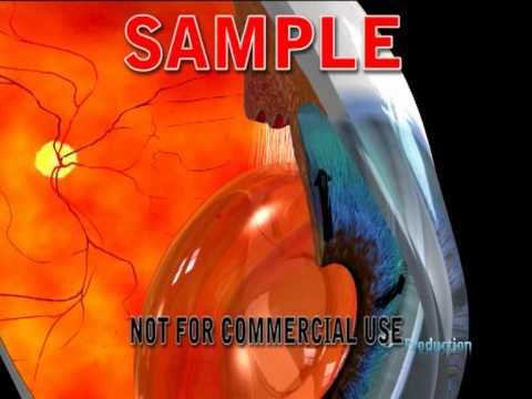
A not-awful 3D review of some basic eye structures.
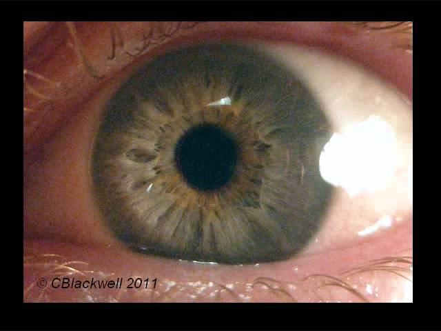
Two great lectures by Dr. Blackwell

Three part video on eye anatomy by Dr. Sousa. I found his lectures pretty good, and relatively all inclusive. There aren’t many videos like this online, oddly enough. Thanks Dr. S!
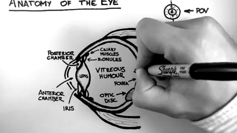
An interesting hand-drawn video on basic eye anatomy.
A “super” lecture explaining the difference between 3rd, 4th, and 6th nerve palsies!
Learn how fire, acids, paintball guns and fireworks damage the eye. This lecture is explosive!

Understanding human eye anatomy by examining the variations found in nature.
A humorous 24-minute guide to the slit-lamp eye exam. Every student needs to see this!
Finally! A good explanation of motility problems and how to measure them.
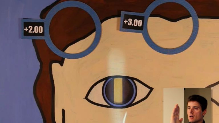
The hardest skill to understand and master. Root makes it easy!
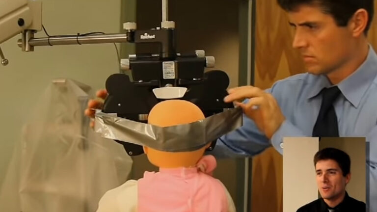
A short and humorous review of the child eye exam.
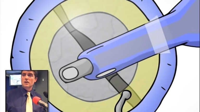
Surgery is explained using a cartoon model, followed by live demonstration.

The history of cataract surgery and how it has advanced.

The basic optics you need to know to understand glasses prescriptions.
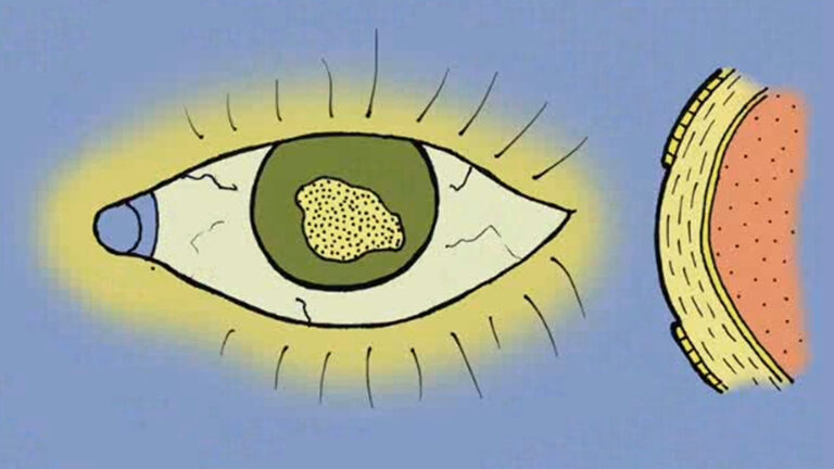
Common eye trauma like corneal abrasions, lid lacerations, and globe rupture.
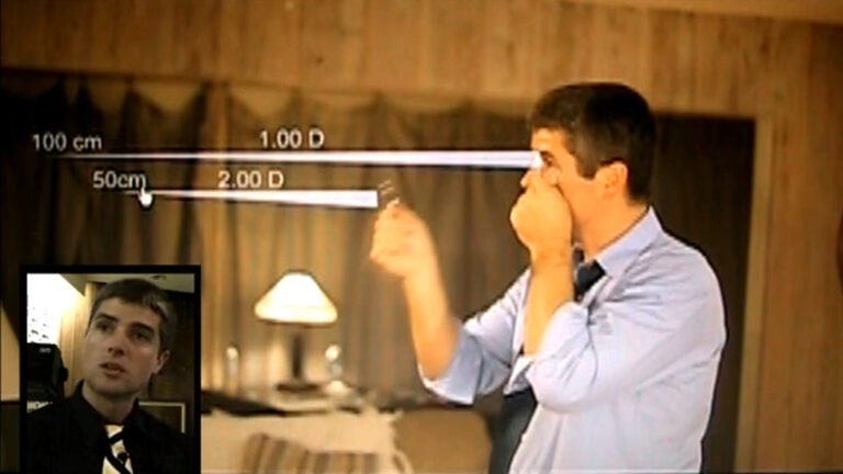
Explaining optics concepts like diopters, astigmatism, and glasses.
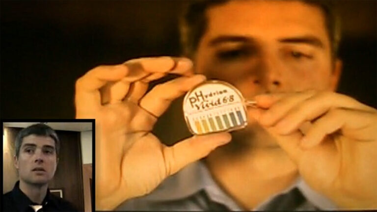
Abrasions, Lacerations, chemical burns, and open globes.

Advanced eye tricks for difficult and challenging patient encounters.
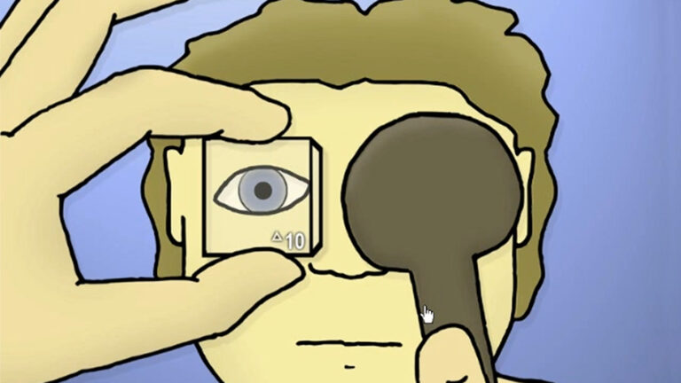
The weird things we check with kid eye exams.
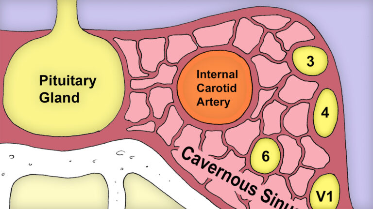
Eye movements, nerve palsies, and localizing neurologic lesions.
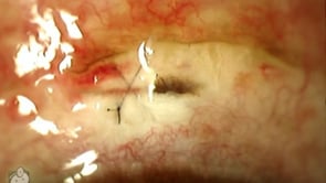
This cataract complication is rarely seen. Here you can see a piece of the iris (the colored part of the eye) that has prolapsed upwards through the cataract incision. This particular surgery was performed with the “scleral tunnel” technique, where the doctor creates the entry incision through the sclera and tunnels into clear cornea before…
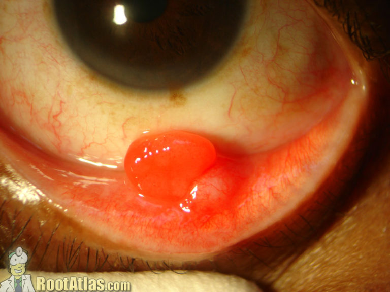
Red bump on inner eyelid, usually from chalazion.

length: 46 seconds This video shows an animatronic eye, designed as a type of art exhibit. The prosthetic is tied into a face tracker. Interesting how we are riveted by the presence of an eye … such that we notice eyes pointed at us, even out of the corner of our vision from across a…
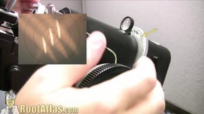
This video shows the use of a lensometer, a device used to check the prescription in glasses. This device can be tricky for the novice technician to use, as you must align the glasses well and move two dials at the same time to hone in on the prescription. Here are the steps: 1. Place…
length: 9:20 minutes Ever wondered how high quality lenses are created? Here’s a great youtube video showing the process. In this case, I believe that these are lenses for high-end cameras, but the process may be similar for our own medical lenses. Seems like a lot of work.

length: 4:40 minutes NOTE: This video has moved. If you want to watch it, you’ll have to go directly to the youtube page here. Some crazy person redid the lyrics to a Justin Timberlake music video and turned it into a glaucoma rap. “Am I going to have to start you on XalatAN, or perhaps…
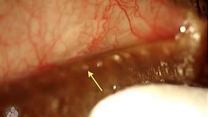
MGD with blocked glands causing blepharitis and dry eye.
length: 30 seconds Another argument for those contact lenses!
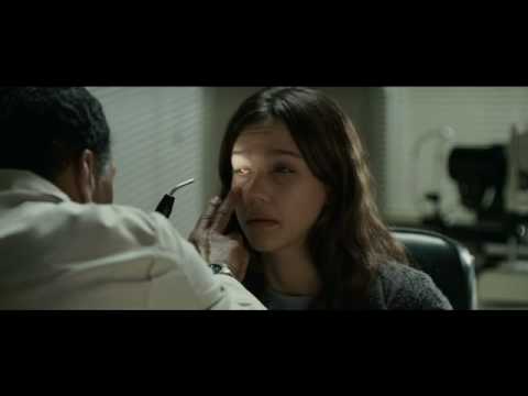
Looks like Jessica Alba is in a movie about a possesed corneal transplant. See if you can notice any ocular inconsistancies in this trailer! Here are the ones I found!! Problem 1. Unrealistic visual expectations The dialogue, and Ms. Alba’s proficiency with her walking cane, seem to indicate that she’s been blind since birth, or…
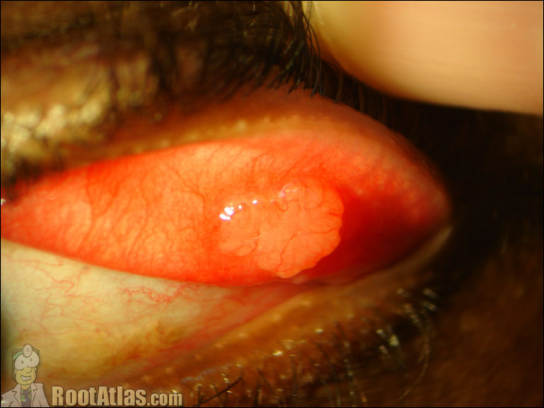
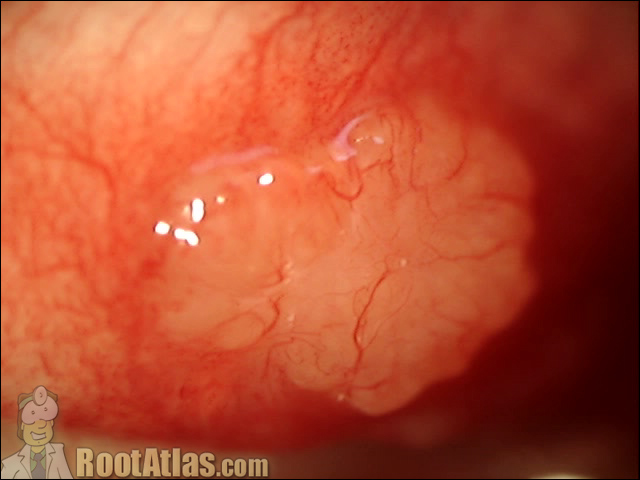
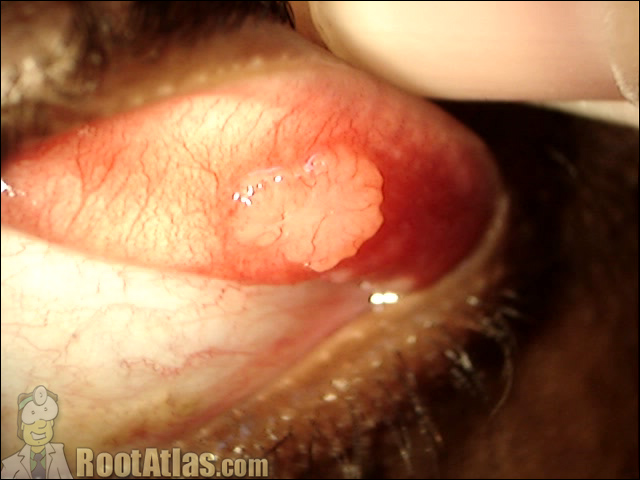
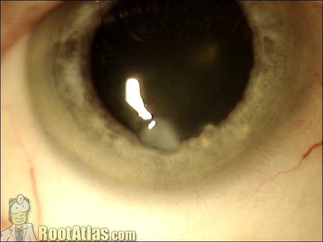
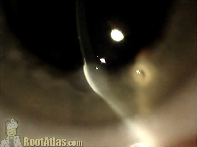
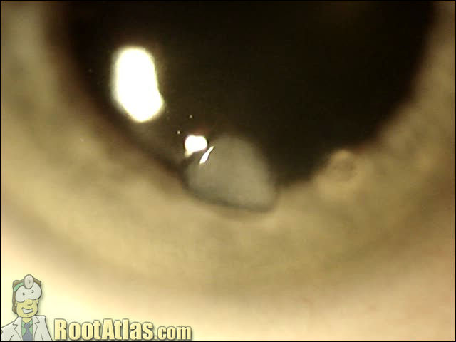
This photograph shows a corneal opacity called a Salzman nodule on the surface of the eye.
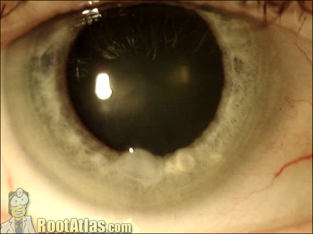
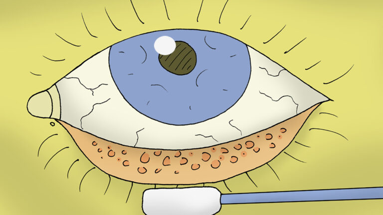
All the eye infections you can stomach.
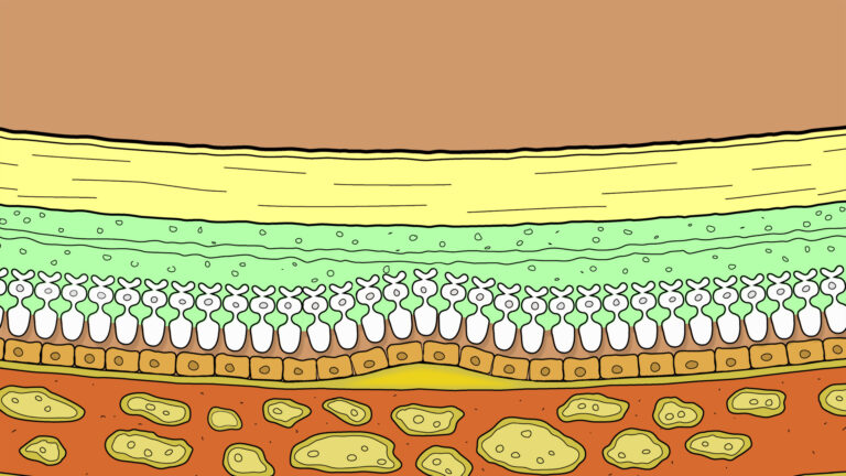
From diabetic retinopathy to detachments.
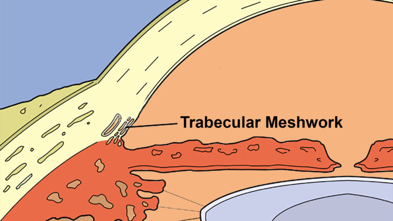
Chronic glaucoma and acute glaucoma.
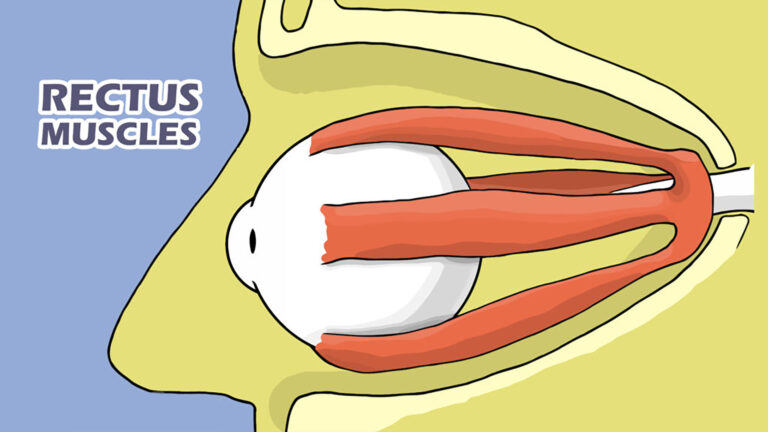
You need to learn the anatomy first to understand the eye.
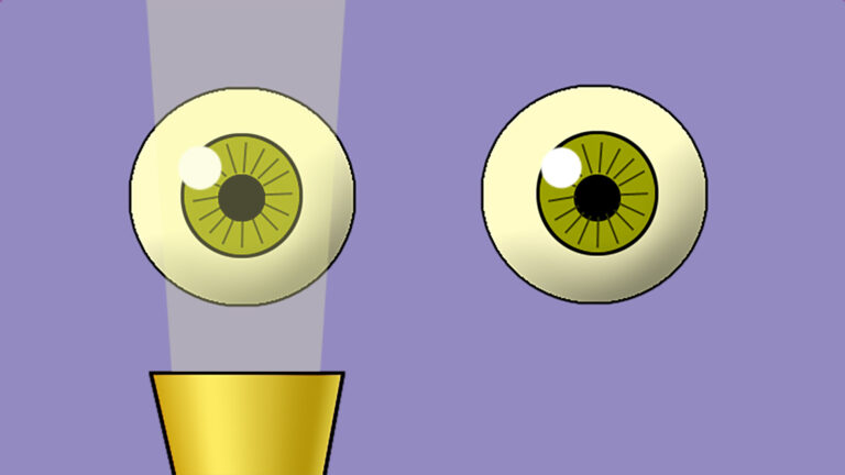
The ocular HPI and exam documentation.
This video shows an eye with significant cornea band keratopathy, and how it is removed with EDTA chelation. About this conditionBand Keratopathy, is a precipitation of calcium salts that forms under the epithelium. It occurs in a band pattern across the middle cornea that is most exposed to air. The believe is that evaporation of…
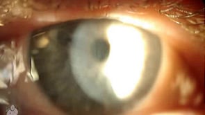
This video shows a cornea with amiodarone verticillata deposits. You can see these as a whorl pattern – the entity is also called whorl keratopathy or hurricane keratopathy. These deposits are benign, difficult to see, and rarely (if ever) have any visual significance. Drugs that can cause this pattern: CACTI Mneumonic: chloroquine, amiodarone, chlorpromazine, tamoxifen,…
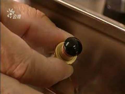
length 4:44 This video shows how a contact lens is made … from lathe, to polisher, to sterilization. The process is more involved than you might think, though disposable lenses must be more automated then what’s seen here. Interesting how topography is used to check the surface of the lens.

length 6:47 This video shows how prosthetic eye inplants are created. Typically, after the eye has healed from an enucleation, the conformer is replaced by the prosthesis by an ocularist. An interesting video on the artistry behind these devices.
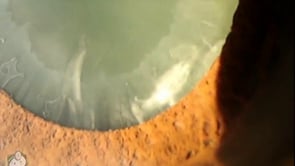
This video shows an eye suffering from severe pseudoexfoliation syndrome of the lens. This has caused glaucoma and will make his cataract surgery difficult. In this movie, you can see the white PXF material on the surface of the lens – it looks radially oriented because the iris rubs against the lens at this point….
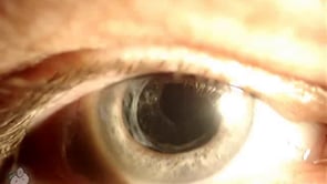
This video shows a cluster of Elschnig pearls that have formed behind the lens after cataract surgery. These grape-like clusters form from residual lens epithelial cells that migrate along the remaining capsular bag. They can be seen with a microscope and are usually visually insignificant. When epithelial cells form in the central visual axis, this…
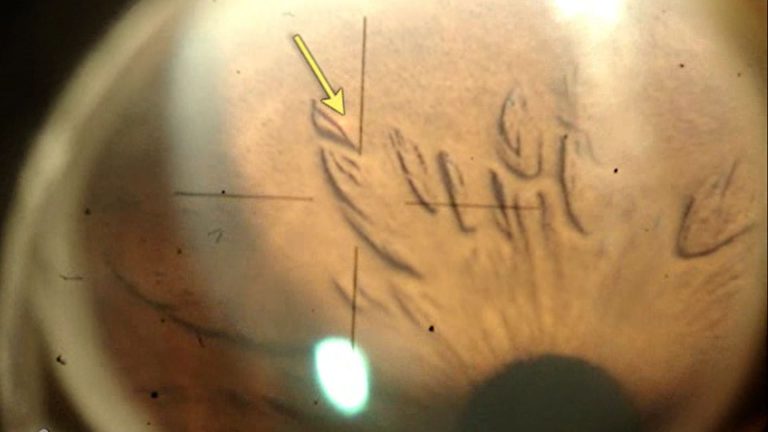
This video shows a laser iridotomy performed on an eye with angle-closure glaucoma. A laser periphery iridotomy (LPI) is a procedure where you use a laser to blast a hole through the iris. This allows fluid from behind the iris to flow forward into the anterior chamber … and eventually drain out of the eye….
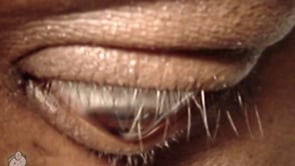
A notch in the lower lid from KCN.
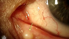
A keratin-filled cyst.
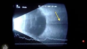
This video shows an ultrasound of the eye. The cornea and lens are on the left portion of the movie, while the retina is on the back surface. The black line coming out the back of the eye is the optic nerve. As the eye moves you can also see some stuff floating in the…
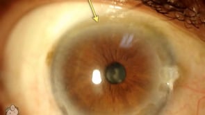
This video shows an eye with Terrien’s Marginal Degeneration. This is when the cornea thins at the edges toward the limbus. The etiology of this condition is not understood, but classically these eyes present with a band of thinning with a step-off toward the center. Also, you can see white lipid deposit, vessels growing into…
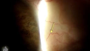
A rhythmic twitching of the eye. It’s subtle.
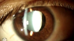
This video shows a cataract lens that has dislocated backwards after blunt trauma. You can even see a veil of vitreous coming around the lens into the anterior chamber. This cataract will be difficult to remove, as an anterior approach is unsafe (there is too much zonular dehiscence such that the lens might fall into…
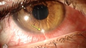
This video shows a sterile corneal infiltrate at the inferior limbus in an eye with blepharitis. This opacity is entirely sterile and occurs from a hypersensitivity reaction at the limbal vessels in the cornea. This eye was treated successfully with good lid hygeine and a mild steroid. This entity is usually bilateral, and as you…
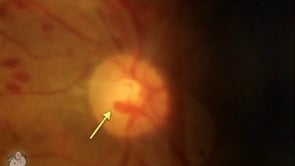
This video shows what spontaneous venous pulsations look like in the retina. This eye is suffering from ocular ischemic syndrome which has dilated the retinal veins and make them even easier to see than usual. Most people believe that these pulsations are caused because of a differential between eye and CSF pressure. The CSF cavity…
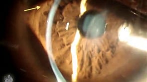
This video shows what the Rizzuti’s light reflex looks like in eyes with keratoconus. You perform this test by shining a penlight into the eye at an oblique angle. The cone shape of the cornea causes the resulting iris light reflection to a point. This is hard to explain, so you should check out the…
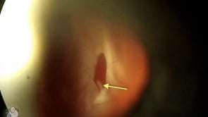
This video shows a retinal detachment of the eye as viewed from the slit-lamp microscope. In this case, you see a large bullous rhegmatogenous detachment with a retinal tear/hole at the 1 o’clock position (the image is reversed). Unless you are experienced with the retinal exam, this movie may be hard to interpret … retina…
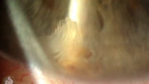
This eye has a small piece of lens nucleus sitting in the bottom of the anterior chamber after successful phaco cataract extraction. This usually occurs when a piece of the hard nucleus gets stuck in the angle and isn’t seen in surgery because of dense arcus or corneal edema (especially sub-incisionally). This nuclear fragment eventually…
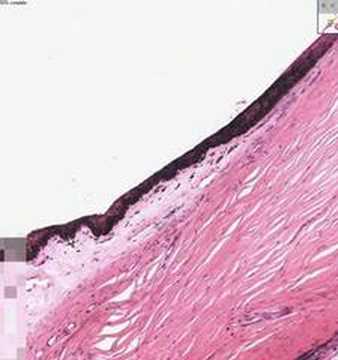
length: 8 minutes This short video goes through basic ocular histology by careful examination of a microscopic slide of the eye. This is a quick and succinct pathology review of the ocular structures from cornea working back to the retina.
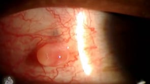
These bumps are normally harmless and react well to steroids.
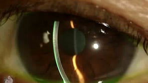
This video demonstrates what cell and flare look like under the slit-lamp microscope. “Cell” is the individual inflammatory cells while “flare” is the foggy appearance given by protein that has leaked from inflamed blood vessels. This finding is commonly seen with uveitis, iritis, and after surgery … and actually seeing it can be challenging for…
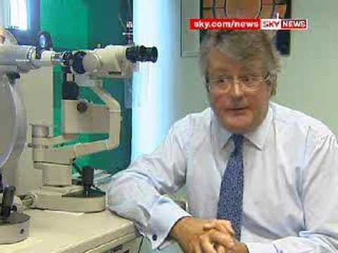
length: 2:23 minutes This video shows the use of night-time hard contact lenses to change the prescription of the eye. This is orthokeratology, and can be used to temporarily mold the cornea into a steeper shape and overcome small amounts of astigmatism and near-sightedness. This method isn’t used often by ophthalmologists in the US, but…

This video is an English news interview about the first injections of for retinitis pigmentosa into a human subject at Moorfields. A review of litereature shows that Dr. Ali has been working on several mouse models of RP and vector delivery. This pubmed link for more info on gene research. This link goes directly to…
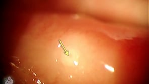
The opening to the nasolacrimal system.
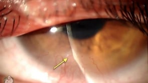
A tiny silicone stopper for the punctal opening.
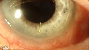
This slitlamp video shows a phylectenule bump on the surface of the cornea. This inflammatory nodule is caused by a delayed hypersensitivity reaction to staph antigens in a case of bad blepharitis. This case was successfully treated with good lid hygeine, erythromycin ointment, and a mild steroid. Download this video To download this video, right…
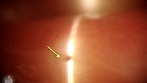
This video shows a perfluoron bubble sitting in the bottom of the eye in the anterior chamber. This chemical is used during retinal surgery to help lay down the retina. Here it has somehow gotten into the front portion of the eye. Since perfluorocarbon fluid is heavier than water, the bubble has sunken to the…
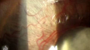
This video shows new, abnormal blood vessels growing on the surface of the iris. This NVI (neovascularization of the iris) is usually seen with bad diabetic retinopathy or central retinal vein occlusions. Any ischemic state in the retina can lead to VEGF upregulation … thus promoting new vessel growth. Iris vessels are an ominous sign,…
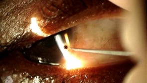
This video shows a small piece of metal stuck to the surface of the eye. It is removed from the cornea using an 18 gauge needle at the slit-lamp microscope. Particles like this need to be removed because they are painful, rust quickly, and can lead to infection and corneal ulcer. Sometimes it is possible…
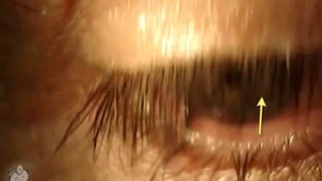
The effect of prostaglandin drops on eyelashes.
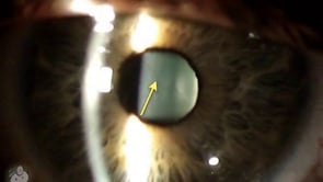
In this video, you can see the normal suture lines that form in the lens with normal eye development. In the front of the lens is a “Y” while in the back, the “Y” is inverted (like a mercedes sign). This is a subtle finding and easier to see in a young eye. Certain types…
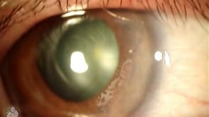
This video shows epithelial ingrowth that occurs sometimes after LASIK surgery. If you look closely at the lasik flap, you can see an area of white deposits in an island configuration. These are collections of corneal epithelial cells that have grown under the flap. The surface epithelial cells can proliferate quickly, explaining why surface abrasions…
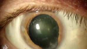
This video shows an iris coloboma defect in the eye. As you can see here, the iris muscle hasn’t completely formed in the infero-nasal portion of the globe. This gives the normally round pupil a “keyhole” appearance. If you look closely, you can even see the edge of the lens … which you can’t normally…
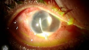
This video shows an eye with a hypopyon. This is when pus layers out in the front portion of the AC and can be seen as white in the bottom portion of this movie. Hypopions can be sterile or infectious. This particular case was secondary to bacterial endophthalmitis from an exposed tube-shunt (a tube placed…
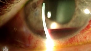
This video shows a layer of blood inside the eye called a hyphema. Hyphemas like this one can occur after blunt injury when the delicate iris arteries bleed into the front anterior chamber of the eyeball. Treatment of hyphemas involves steroid drops (to decrease inflammation) and cycloplegics (to dilate the eye for comfort and to…
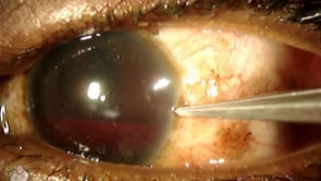
This video shows an eye with a large hyphema (blood) inside the anterior chamber. The blood has settled into the bottom 35% of the eye, while the upper portion of the AC is dense with free-floating RBCs. In the last half of the movie, I create a paracentesis using a super-blade scalpel. Hyphemas typically occur…
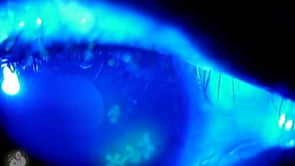
This video shows the classic dendritic ulcer seen with HSV keratitis. You can see them easily with the use of fluorescein staining (that’s why everything is blue in the movie). These patients typically present with a painful, red eye with photophobia. The HSV is typically the type-1 hsv infection that most people are inoculated with…
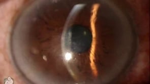
This video shows a cornea with Fuch’s dystrophy. You can see guttata or guttae on the back surface of the cornea. These bumps indicate endothelial pump difficulty, and appear as a “beaten metal” appearance. If you look closely (look where the arrow is pointing) you can see a pock-marked surface that looks like craters on…
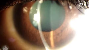
In this video you can see particles stuck to the back surface of the cornea. These are KP (keratic precipitates) which are clusters of inflammatory cells that tend to congregate and stick to the endothelium during times of AC inflammation (uveitis). As you can see in this video, the KP spots are visible on the…
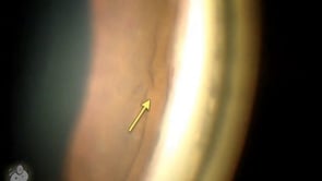
This gonioscopy video shows an interesting finding: the blue line is a haptic from an intraocular lens that has poked through the iris into the anterior chamber. Fortunately, the eye pressure was not affected, and there does not appear to be any inflammatory response (ie. you don’t see any PAS in this video). Gonioscopy is…
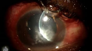
This slit-lamp video shows a large, central corneal ulcer that was caused by fusarium fungus. Classically, fungal ulcers are described as gray, feathery borders, with satellite lesions. In reality, the only way to determine the real pathologic cause is with a good culture (molds take forever to grow and require a deep sample) and time…
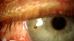
This video shows the removal of metal particle from the surface of the eye using a needle. Metal particles can occur from metal grinding and machine work … the metallic flakes have a tendency to get into hair then fall onto the cornea and stick. This quickly rusts and if not removed soon, can cause…
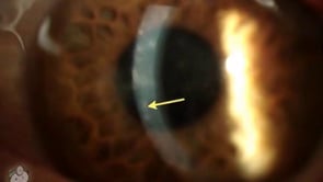
This video shows an eye with EKC viral conjunctivitis, caused by the adenovirus. Here you can see numerous subepithelial infiltrates. The easiest way to visualize these is by aiming your slit-lamp light so it comes in from the side and diffusely illuminates the corneal surface. This cornea is suffering from EKC (epidemic keratoconjunctivitis). The adenovirus…
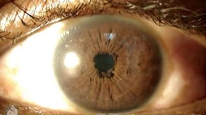
This video shows what appears to be iris sphincter hypertrophy at the pupil margin. Some might also describe this as very mild ectropion uvea. Download this video To download this video, right click on a link below and choose “Save Target As…” ectropionuvea.wmv (3.0 meg, Windows video file) Screencaptures
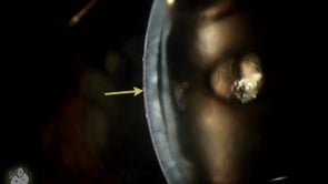
This eye has undergone DSEAK (Descemet’s Stripping Automated Endothelial Keratoplasty) also called DSEK. As you can see here, the endothelial transplant has separated from the inner cornea. This was reopposed with sf6 gas in the anterior chamber. DSEK has many advantages over traditional PK and is a wonderful alternative procedure for certain conditions. Download this…
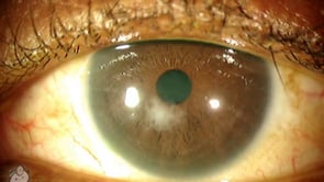
This eye has had Lasik, and under the corneal flap you can see a small scar. This resulted from DLK (diffuse lamellar keratitis) that can occur when debris at the flap interface causes a non-infectious inflammatory response. This condition is also called the Sands of the Sahara because of the usual wavy appearance of the…
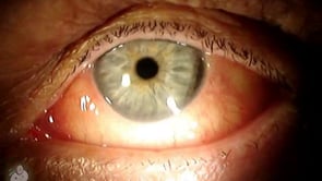
This cornea has a small dellen (thinning of the corneal stroma) that occured after cataract surgery. The cornea thins when it is dehydrated, and in this case, a localized area of conjunctival edema next to the limbus interfered with the normal tear-blink mechanism. Fortunately, this thinning quickly resolved with aggresive lubrication. Download this video To…
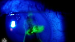
This video shows a cornea with a small laceration through the stroma. You can tell that the wound goes all the way into the anterior chamber as the wound is seidel positive when fluorescein is applied. Download this video To download this video, right click on a link below and choose “Save Target As…” cornealaceration.wmv…
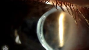
This video shows an intra-stromal lens that’s been inserted directly into the cornea. These lenses are no longer used as they caused many problems with corneal oxygenation. Download this video To download this video, right click on a link below and choose “Save Target As…” corneallens.wmv (5.3 meg, Windows video file) Screencaptures
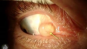
Harmless (but annoying) fluid filled cyst.
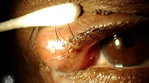
Pushing on a friend’s chalazion.
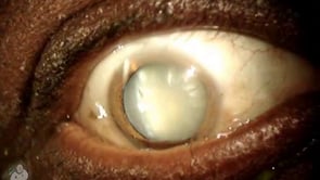
This video shows a dense cataract in the eye. This lens has opacified to the point that it can be seen without a microscope, and is severely limiting vision. We don’t see many cataracts like this in the U.S. as most people have surgery long before. Download this video To download this video, right click…
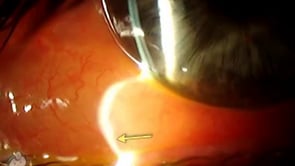
Impressive swelling from allergic inflammation.
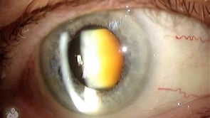
In this video you can see what a typical cataract looks like under the microscope in real life. The central nucleus has turned brunescent (yellowish) such that this patient is suffering from decreased vision that is exacerbated by bright lights. The lens is constructed with three components and has a configuration similar to a “peanut…
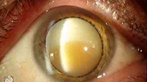
This video shows a hypermature morgagnian cataract. As you can see, the cortex has liquefied and turned milky, while the central nucleus has turned brown and sunken to the bottom of the capsular bag. Download this video To download this video, right click on a link below and choose “Save Target As…” hypermaturecataract.wmv (3.6meg, Windows…
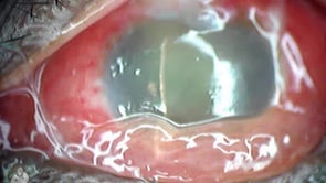
A nasty infection years after glaucoma surgery.
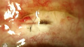
Seidel positive leak.

This video shows the view you have when checking eye pressure (glaucoma check) with the goldman applanation tonometer. This device flattens the corneal surface of the eye, and by determining the amount of force required to flatten a measured area, can calculate the inner ocular pressure. Applanation is one of the harder steps for the…
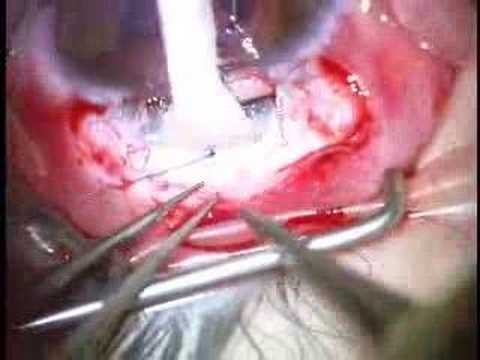
This video shows the surgical technique for performing the glaucoma trabeculotomy surgery. This procedure involves cannulating Schlemm’s canal externally and ripping into the anterior chamber to improve overall drainage. This treatment is primarily done for primary congenital glaucoma, especially with corneal clouding that would preclude goniotomy. In this video, the surgeon creates a square flap…
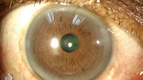
This video shows an anterior polar cataract sitting on the front surface of the lens. The forward placement of these opacities cause less visual disturbance than you might think from it’s appearance. Download this video To download this video, right click on a link below and choose “Save Target As…” anteriorpolarcataract.wmv (2.5meg, Windows video file)…
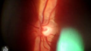
This video was taken at the slit-lamp, and shows what the retina looks like using a hand-held 90 diopter lens. The view is less magnified than the direct scope, and allows viewing of the entire optic nerve and posterior pole. Here you can see both the “cup” (the inner divot) and “disk” (the entire round…
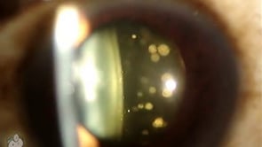
Asteroid hyalosis describes white floaters in the vitreous humor of the eye. These bodies are different than the typical floater, and usually don’t cause visual disturbances. They are caused by calcium soap deposits and can be associated diabetes, hypertension, and high cholesterol. Though usually visually benign, these rarely cause significant visual disturbance. The opacity usually…
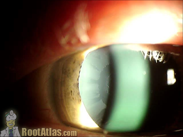
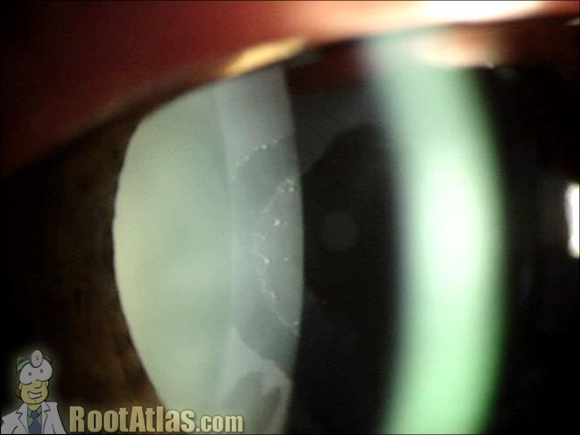
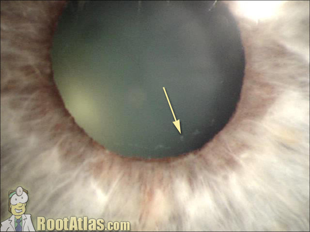
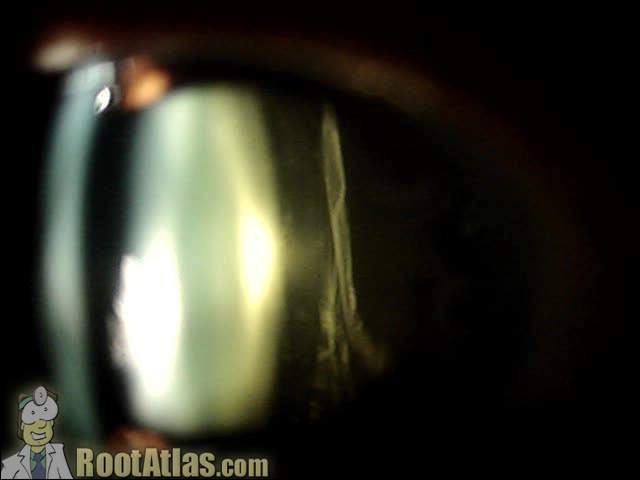
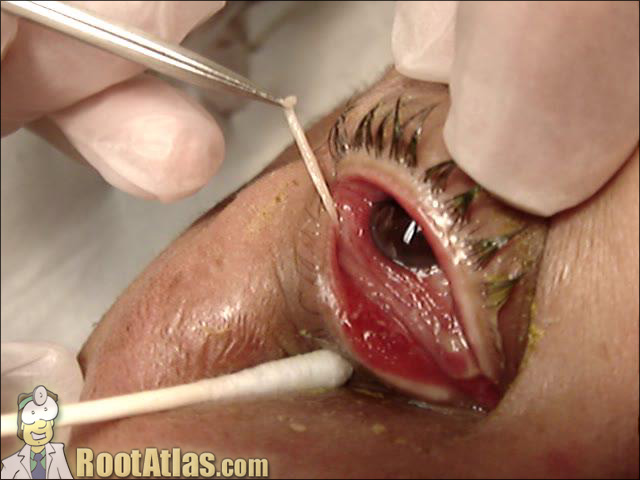
e-eye.thumbnail.jpg” alt=”pseudomembrane-eye.jpg” />
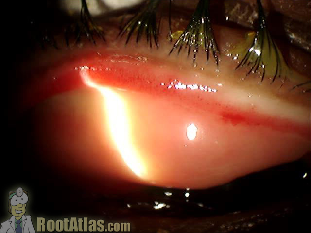
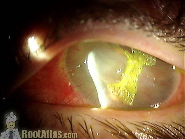
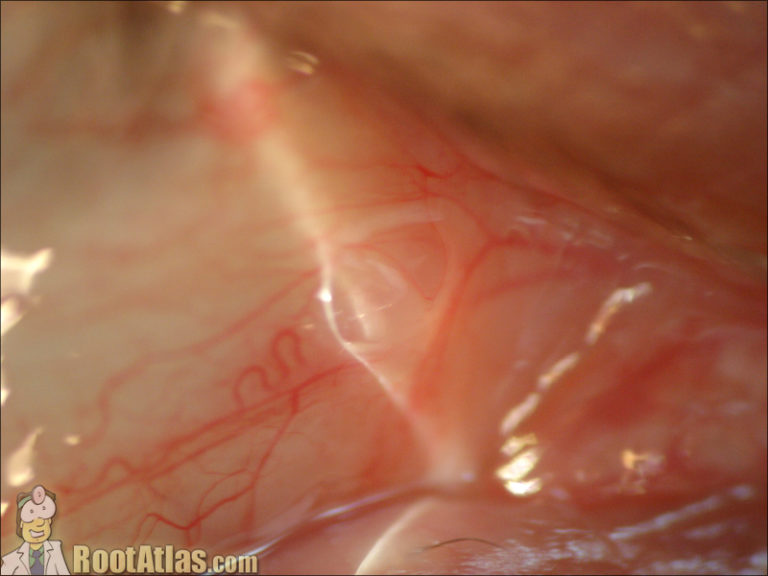
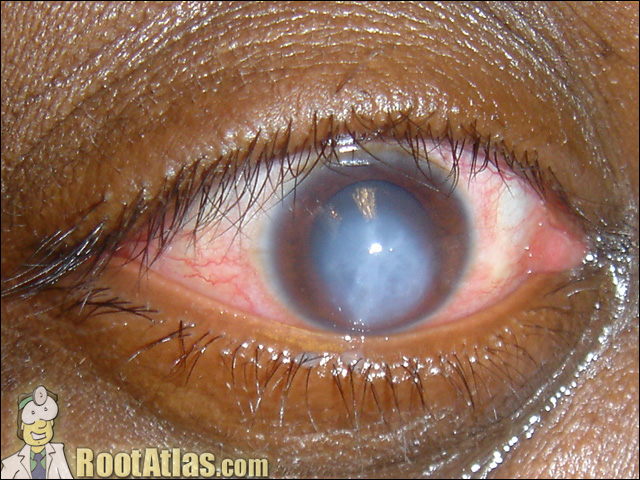
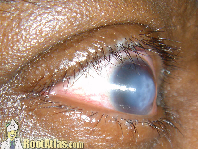
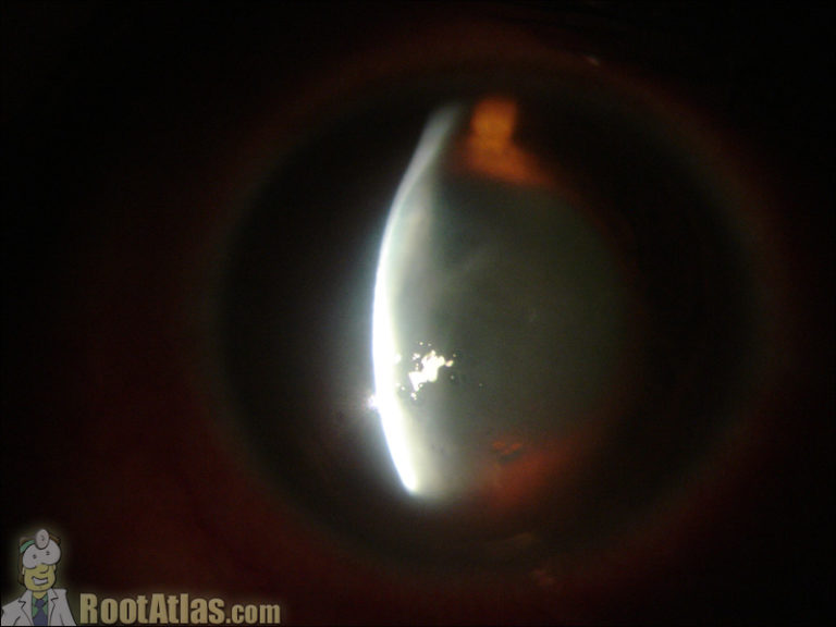
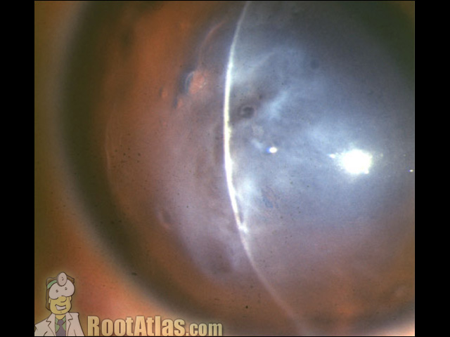
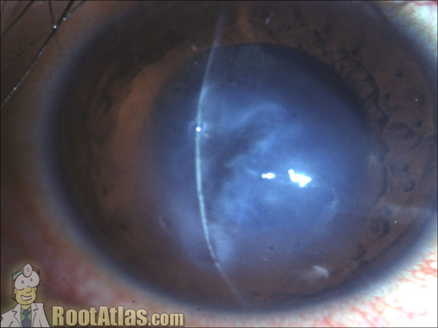
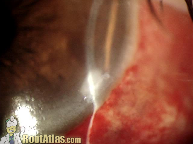
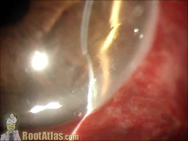
This photo shows corneal thinning caused by a dellen (local dehydration of the cornea). Despite this “thinning” none of the cornea has been lost, it’s just dessicated. These occur from excessive localized drying. In this case, a large spontanoues conjunctival hemorrhage has formed under the conjunctiva. The conjunctiva is epecially elevated right at the limbus…
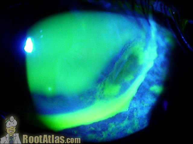
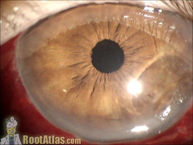
This photo shows a dellen (thinning of the cornea) that occurs from dehydration. You can see it at the limbus in the bottom-right of this picture. You can also see the funny reflection it causes on the iris underneath. Dellen occur when the tear film does not cover the eye. In this case because of…
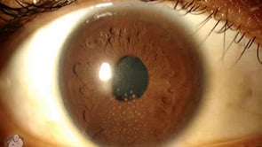
This video shows keratic precipitates that have formed on the back surface of the cornea. These deposits occur from inflammation. Download this video To download this video, right click on a link below and choose “Save Target As…” granulomatouskp.wmv (2.5meg, Windows video file) Screencaptures
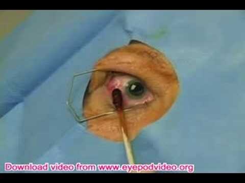
length 3:06 About this Video A pretty nice explanation of the intravitreal avastin, anti-VEGF injection.
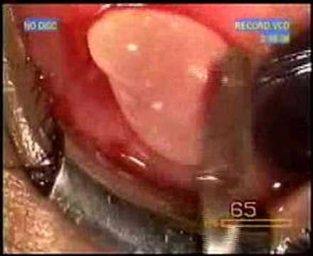
length 2:00 About this Video Removal of a cyst from the conjunctiva. Totally weird.
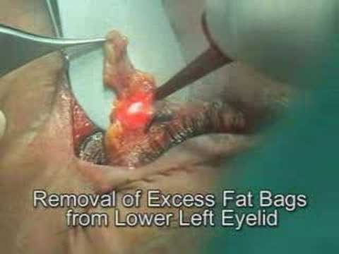
About this Video In this video the CO2 laser is used …
About this Video This is a brief news segment on the treatment of keratoconus using corneal implants and riboflavin collagen crosslinking. I believe C3R is simply riboflavin with a few other chemicals added (patentable?). I’ve been using simple riboflavin like in the original studies. More info on the doctor cited: http://www.keratoconusinserts.com/cornea.html
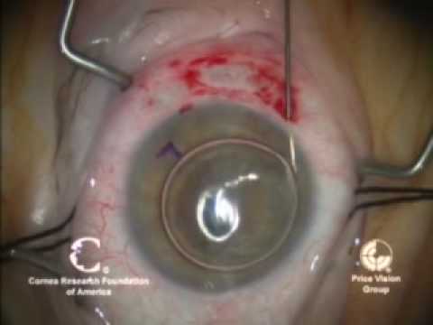
About this Video Descemet’s Stripping Automated Endothelial Keratoplasty. Very well narrated.
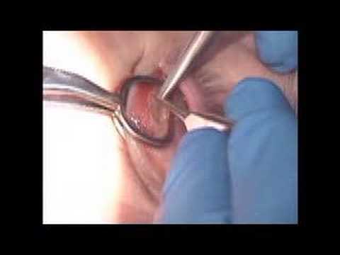
About this Video info
About this Video Info …
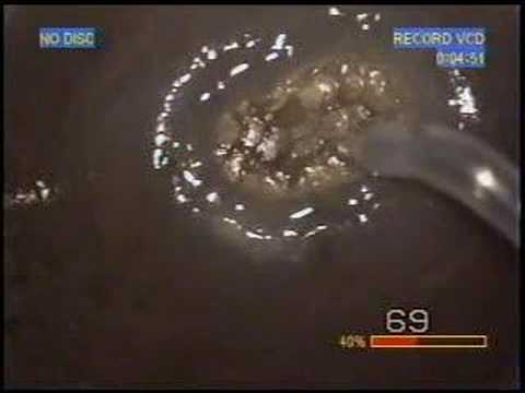


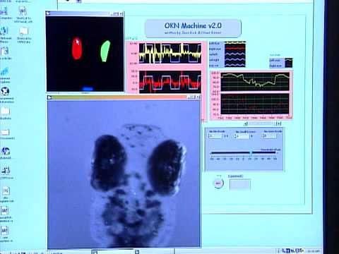
About this Video The zebrafish is a popular aquarium fish with stripes running down it side. Recently, it has become a new model for ocular diseases like RP. This video is an interesting look at …
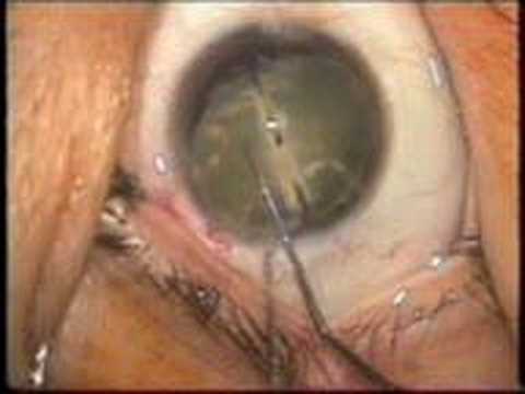
About this video Info
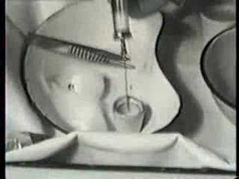
About this video This video shows the amazing effect that prostigmin…
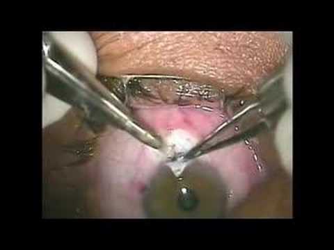
Editor’s Coments: This trabeculectomy is performed with a conjunctival approach. Nice review for a resident or if you haven’t done one in a while. Enjoy.

In this video, Dr. Mizen speaks about some of the ocular manifestations of Myasthenia Gravis and the eye complaints that patients may present to you complaining about. Dr. Mizen speaks about the ocular manifestations of myasthenia Gravis. Another video on myasthenia. This one seems to be at the same event as this other video. The…