Iris | Slit Lamp Video | Video
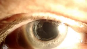
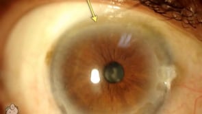
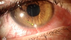
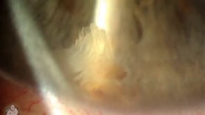
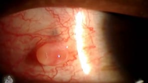
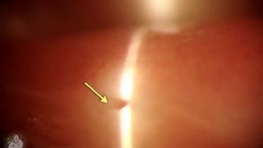
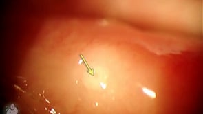
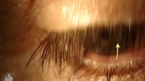
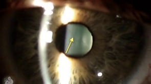
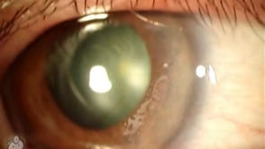
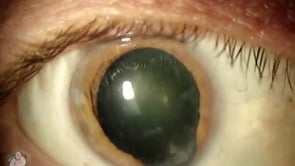
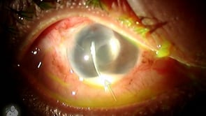
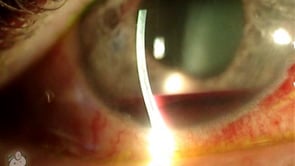
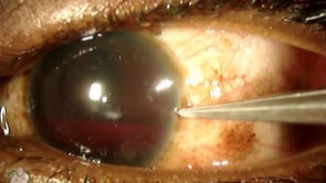
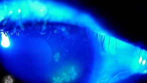
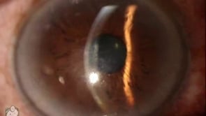
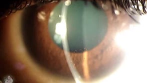
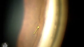
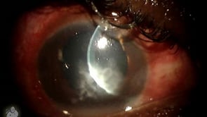
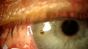
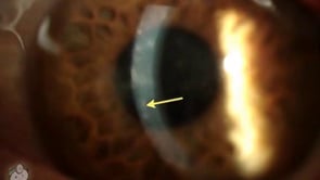
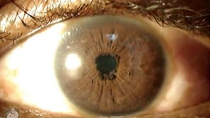
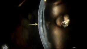
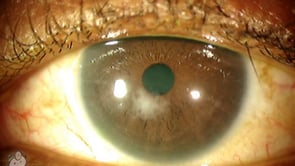
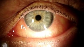
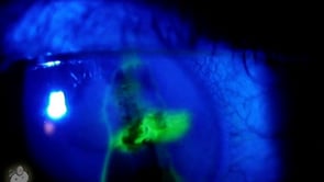
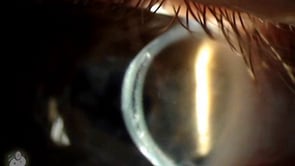
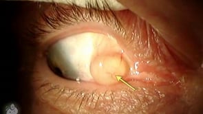
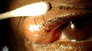
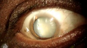
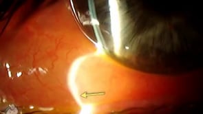
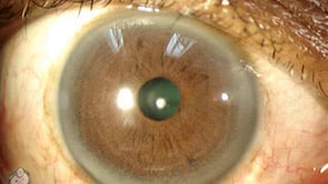
Iris Prolapse after Cataract Surgery (Video)
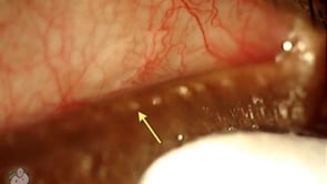
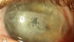
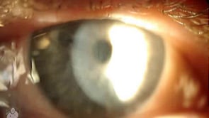
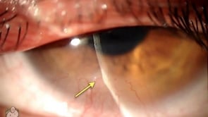
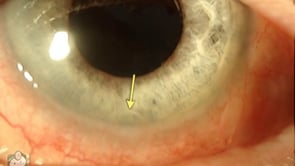
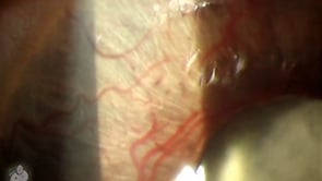
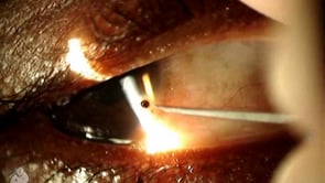
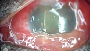
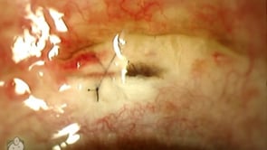
This cataract complication is rarely seen. Here you can see a piece of the iris (the colored part of the eye) that has prolapsed upwards through the cataract incision. This particular surgery was performed with the “scleral tunnel” technique, where the doctor creates the entry incision through the sclera and tunnels into clear cornea before…