Anterior Chamber

Retained lens material after cataract surgery (Video)
| |

Retained lens material after cataract surgery (Video)

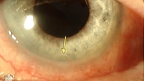
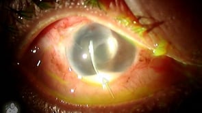
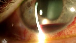
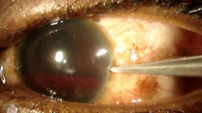
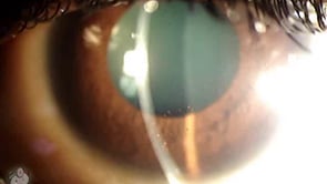
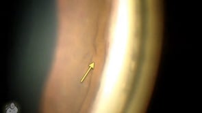
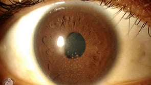
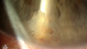
This eye has a small piece of lens nucleus sitting in the bottom of the anterior chamber after successful phaco cataract extraction. This usually occurs when a piece of the hard nucleus gets stuck in the angle and isn’t seen in surgery because of dense arcus or corneal edema (especially sub-incisionally). This nuclear fragment eventually…

“Cell and flare” in the eye (Video)
| |

“Cell and flare” in the eye (Video)

This video demonstrates what cell and flare look like under the slit-lamp microscope. “Cell” is the individual inflammatory cells while “flare” is the foggy appearance given by protein that has leaked from inflamed blood vessels. This finding is commonly seen with uveitis, iritis, and after surgery … and actually seeing it can be challenging for…

KP (keratic precipitates) on the back of the cornea (Video)
| |

KP (keratic precipitates) on the back of the cornea (Video)

In this video you can see particles stuck to the back surface of the cornea. These are KP (keratic precipitates) which are clusters of inflammatory cells that tend to congregate and stick to the endothelium during times of AC inflammation (uveitis). As you can see in this video, the KP spots are visible on the…