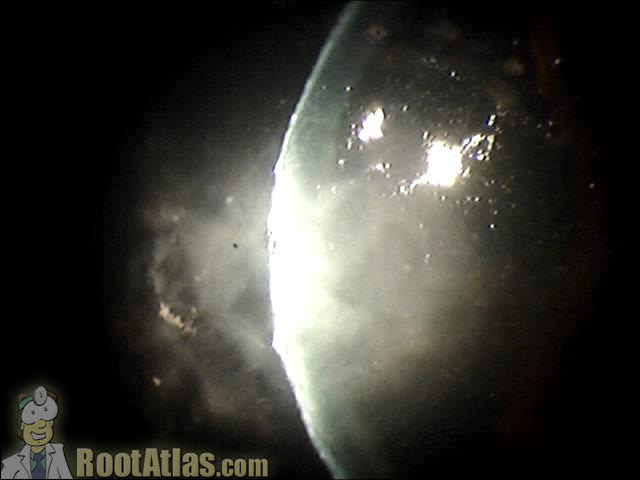
Fungal corneal ulcer (Video)
This slit-lamp video shows a large, central corneal ulcer that was caused by fusarium fungus. Classically, fungal ulcers are described as gray, feathery borders, with satellite lesions. In reality, the only way to determine the real pathologic cause is with a good culture (molds take forever to grow and require a deep sample) and time course.
Fungal ulcers like this are difficult to treat and resolve slowly. The general medical therapy is topical amphotericin and oral/iv systemic coverage with fluconazole or voriconozole.
Download this video
To download this video, right click on a link below and choose “Save Target As…”
fungalcorneahypopion.wmv (5.6 meg, Windows video file)
Screencaptures


Good job!! Commendable
nice job bt i wish it would b more clear………..
Goodevening…
Please,if you can,send to me to my e-mail all your educational ophthalmology videos.
thanks
good &informative video, thanks
excellent work.
plz send to me to my email all of your educational videoes thanks and so nice of you for this anticipation
thanks for these clear and inerristive videos