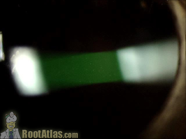
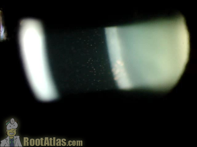
“Cell and flare” in the eye (Video)
This video demonstrates what cell and flare look like under the slit-lamp microscope. “Cell” is the individual inflammatory cells while “flare” is the foggy appearance given by protein that has leaked from inflamed blood vessels. This finding is commonly seen with uveitis, iritis, and after surgery … and actually seeing it can be challenging for the beginning ophthalmology residents.
The technique for seeing inflammation is to shorten your light beam, widen it slightly, and angle your light-path such that the beam hits the cornea on the left, the iris on the right, with you focusing on the anterior chamber in the middle of the eye. This allows you to use the pupil as a black background.
The first eye in this movie shows copious pigment floating in AC after a laser iridotomy. You can’t miss the pigment cells floating there. The second segment shows mild inflammatory cells, but a lot of flare (it looks like the beam a film-projector would make in a smokey movie theater). The last segment shows a moderate amount of cell that is moving by convection currents: the cells in the back float upward because the chamber is warmer there, and sink in the front where the aqueous is cooler.
Download this video
To download this video, right click on a link below and choose “Save Target As…”
cellandflare.wmv (8.4 meg, Windows video file)
Screenshots


Well, all comments about iridotomies aside, this is a great video. I am going to use it in a presentation to our residents.
Out of curiosity, Dorri, were your iridotomies done by an ophthalmologist or an optometrist? Some states allow optoms to do them and it seems like everyone and their mother are getting one. Regardless, it’s a shame you weren’t put on steroids. Undoubtedly, you developed an immune response to the uveal tissue floating around in your eye, and your other problems stem from the resultant uveitis.
great video! May I know the details of the digital slit lamp used to produce it?
Just wanted to say thank you for this video, it was very helpful…It will strengthen my ability to perform a more complete eye exam
Thanks for this video!!
It has helped me understand cells and flare much better!
I searched hi and low for such a good explanation.
thanks !!!!!
Thanks for video…very nice video..
wonderfull video. have been looking for some educational videos like these.thanks.
VERY VERY EDUCATIVE VIDEO WHICH GAVE A RICH IDEA AS TO WHAT IS TO BE SEEN IN CASE OF EYE FLARE.
THANK YOU VERY MUCH FOR THIS WONDERFUL STUFF.
Both flare and cells are so small. How do you tell the difference?
I fail to see how a side effect is more likely with an OD vs an MD.
Great videos thanks for this site. Very rich. Have been looking for details like this for a while. Thanks again.
I am a doctor of optometry student (O.D. program) in my 2nd year and really enjoy and appreciate your site. I am planning on doing a post-doctoral optometric residency in ocular disease (1 yr) followed by a glaucoma research fellowship (2 yrs). I will be writing topical Beta blockers, prostaglandins, etc…all day long lol… Your site is a great “brush -up” for me and I really appreciate it! Keep up the good work….
Amazing work. Great videos. Trust me this is a great service. Its so selfless ( allowing to download the videos). God bless you.
Thanks!
Tank you for a good video,I’m sure this video will help me in my job.
Tim,
How can I contact you to ask about using some of your excellent material in a publication I am working on?
Thanks,
Kevin
This video is great, helped me understand cells and flare so much better, thank you 🙂
What is difference between cell and flare?
fantastic video with important tacts how to see flair & ac cells .
very useful.
dr sunil bhujbal
Great effort to make clear flare and cell..
I may not understand if i didnt watch this video..
Sir great work. …you deserve noble prize in science of ophthalmology
Nice video.
Very much instructive vedio, I’ve gathered knowledge about cells and flare. Heartfelt thanks
Great video, thank you so much
This video is so good and I learn to differentiate between cells and flare. Nice one…..
Wow! This video was so clear and concise! Thank you 🙂
nice video to understand about cells & flare specially in active case of uveitis
Best explanation I have ever seen! Thank you.
What should be the optimum magnification and illumination ?
Very nice 👍
superb video..thanks Dr
Omg finally I get it! Thank you 🙂
Recently I was diagnosed with uveitis and as the analysis was being done I was given a running description of what they could see through their slit lamp. I think this video is terrific and should be shown to anyone with that condition so they can have a better understanding of their condition.
Thanks for educating all of us! This is so informative and as most people have quiet ACs its often hard to know what I’m looking for sometimes, so this definitely helped heaps!
Your efforts to create this video and make it so SHORT so people will watch it, Will probably save countless people’s vision! We THANK people like you Tim Root for helping people like us! WELL DONE!!! 👏👍
I work in rural Minnesota ER’s all over the state, and I can tell you, this video will make a HUGE difference in these wonderful rural Minnesota farmers and their families lives!