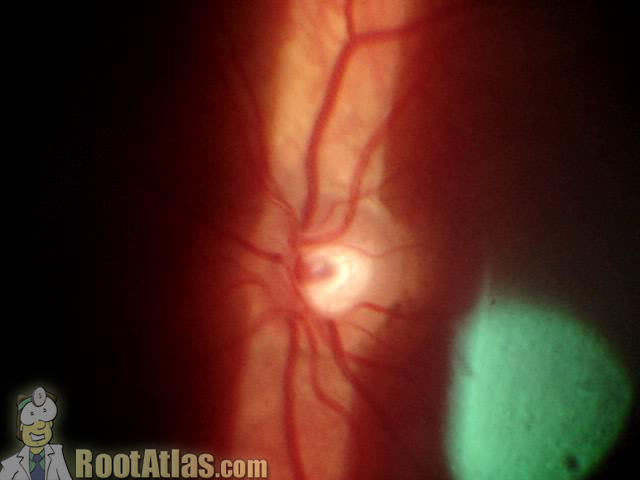
90-diopter view of the retina (Video)
This video was taken at the slit-lamp, and shows what the retina looks like using a hand-held 90 diopter lens. The view is less magnified than the direct scope, and allows viewing of the entire optic nerve and posterior pole. Here you can see both the “cup” (the inner divot) and “disk” (the entire round optic nerve). The movement of the slit-beam gives a stereoscopic sense of depth.
Download this video
To download this video, right click on a link below and choose “Save Target As…”
90diopterretina.wmv (2.5meg, Windows video file)
Screencaptures

Great video and fantastic website. What equipment was used to capture this 90D fundus video?
Thanks for the comment. This video, like many on this site, was simply a consumer digital camera (set on video mode) held up to the eyepiece at the slit-lamp.
Only rarely can I capture good retina video … the lens has to be clear, and the patient tolerate a fair amount of illumination without blinking.
Thanks!!
I like this website!
Great Website, esp. for the young ophthalmologists in the developing world.
Excellent videos, I am really amazed of the quality obtained with just a consumer digital camera. Incredible I have a firewire Camera mounted in my slit lamp through beamspliter, but I am not able to obtain such a great videos. Besides, consider the camera is just help up to the eyepice, what a master in the use of the slit lamp. Congratulations from Ecuador, South America.
I can not see the videos. I see only a green screen and can hear the audio.
Anyone else having the same problem?
Love this site very usefull, thank you.
Fantastic videos really useful for an Optom student in UK
Goodevening…
Please,if you can,send to me to my e-mail all your educational ophthalmology videos for teaching.
thanks
gave me a better idea about what my doctor has been talking about. Thanks
Thanks I liked it very much!