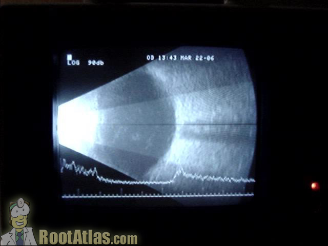
Ultrasound of the eye (Video)
This video shows an ultrasound of the eye. The cornea and lens are on the left portion of the movie, while the retina is on the back surface. The black line coming out the back of the eye is the optic nerve. As the eye moves you can also see some stuff floating in the middle of the image: this is likely just vitreous debris.
When performing an ultrasound, the goal is to image the transducer such that you get a good cut through the optic nerve. When looking for retinal detachments, the retina has a bright reflection line that will spike on the a-scan component.
Download this video
To download this video, right click on a link below and choose “Save Target As…”
ultrasoundnormaleye.wmv (5.2 meg, Windows video file)
Screencapture from this video:

hi could you do a video of the real anatomy of the vitreous body and explain or talk us through it please?
hi!
would just like to know, where was the white line pointed? vertically? or horizontally? i keep getting confused with an axial view and the other type of view.
thanks!
Many thanks for the great videos. I run an ophthalmic diagnostic center and a teaching center. They are most helpful. Would you like a few more on Diagnostic B scan? Fluorescein Angiography/ OCT? I would be happy to contribute
Denice Barsness, CRA, COMT, ROUB, CDOS
CPMC Dept of Ophthalmology
San Francisco