Retinal detachment viewed from the slitlamp (Video)
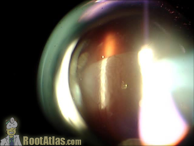
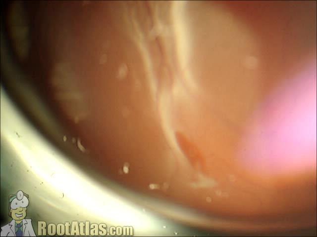
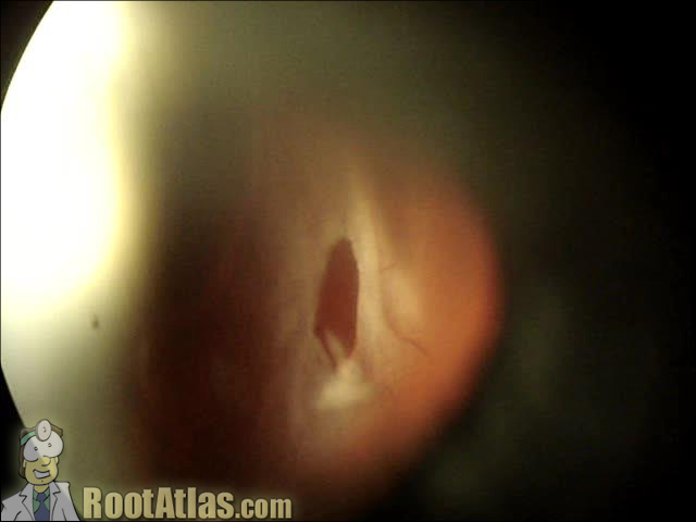
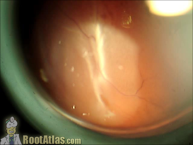
This video shows a retinal detachment of the eye as viewed from the slit-lamp microscope. In this case, you see a large bullous rhegmatogenous detachment with a retinal tear/hole at the 1 o’clock position (the image is reversed).
Unless you are experienced with the retinal exam, this movie may be hard to interpret … retina exam is difficult, and it is even MORE challenging to capture on video. To help you understand this case, I speak through the entire video and label each of the structures.
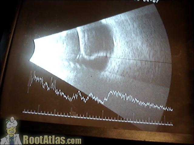
The first third of the movie shows the bullous detachment. The second portion shows the retinal hole, both with a rodenstock contact lens and via a three-mirror lens. The final segment shows the ultrasound for this eye, which looks striking, as you can see how the retina is attached only at the optic nerve and ora serrata. You can also appreciate the retinal spike on the a-scan component of the ultrasound video.
Download this video
To download this video, right click on a link below and choose “Save Target As…”
retinaldetachment.wmv (7.5 meg, Windows video file)
Screenshots
nice video . .
please i would like other related videos to help me in my studies. I am an optometry student studying at KNUST. KUMASI. GHANA
Thank you very much. Helped me appreciate the liquefaction of the vitreous and get a clearer mental picture of it. Great video!
really a very nice vidio it helped me alot
Brilliant video, thanks for sharing
verygood
It’s amazing and help me a lot,thanks for your sharing
great– thank u
Thank you so much for the awesome video! You must spend a lot of time making this video, and it is very worthy because you do teach Med students a lot!
Excellent videos and presentation.