Introduction to Glaucoma Video
This introductory video covers the basics such as open-angle and closed-angle glaucoma. In addition, treatment options, pressure-concepts, and exam findings are shown with full-motion video clips. Overall, I think you’ll find this lecture entertaining.
Screencaptures from this Video:
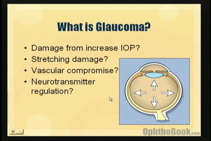
This video begins with a definition of glaucoma … unfortunately, there are many proposed mechanisms and we don’t truly understand this disease.
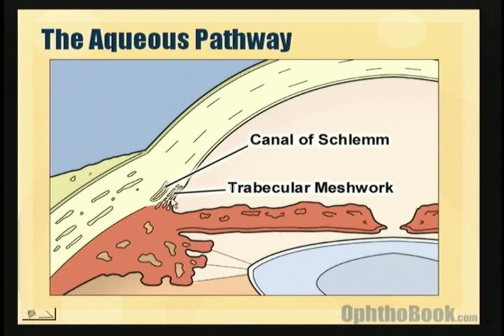
To understand glaucoma, you must understand how aqueous fluid is produced and drained from the eye.
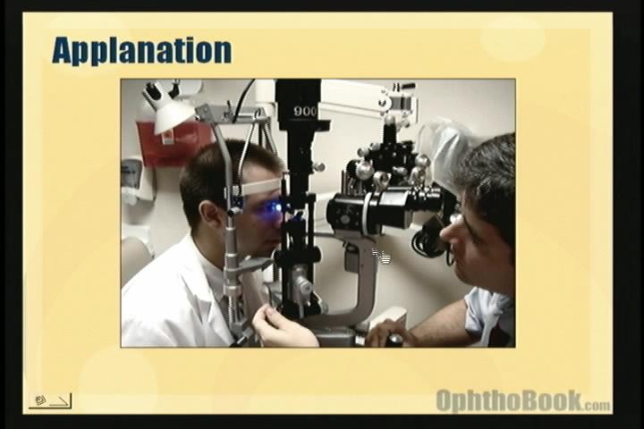
One method of pressure measurement is with the applanation tonometer built into the slit-lamp microscope.
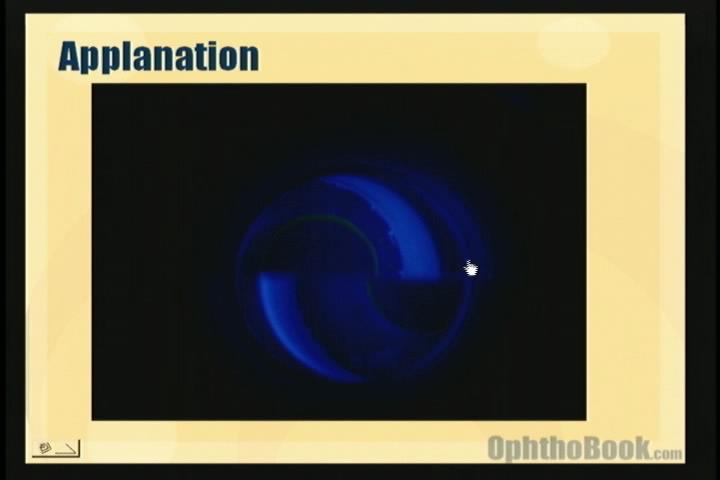
The applanation mires should touch – this lets you know that you’re flattening the proper amount of cornea.
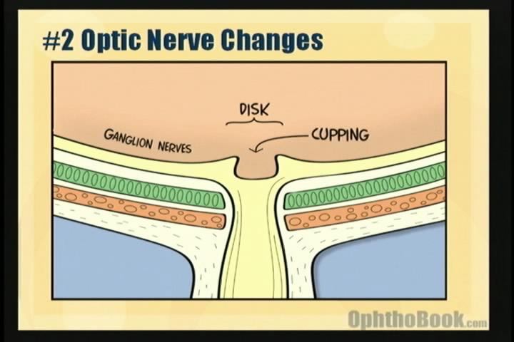
The cup-to-disk area increases as less ganglion nerves are traveling through the optic-nerve.
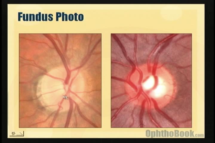
Small cup, and bigger cup.
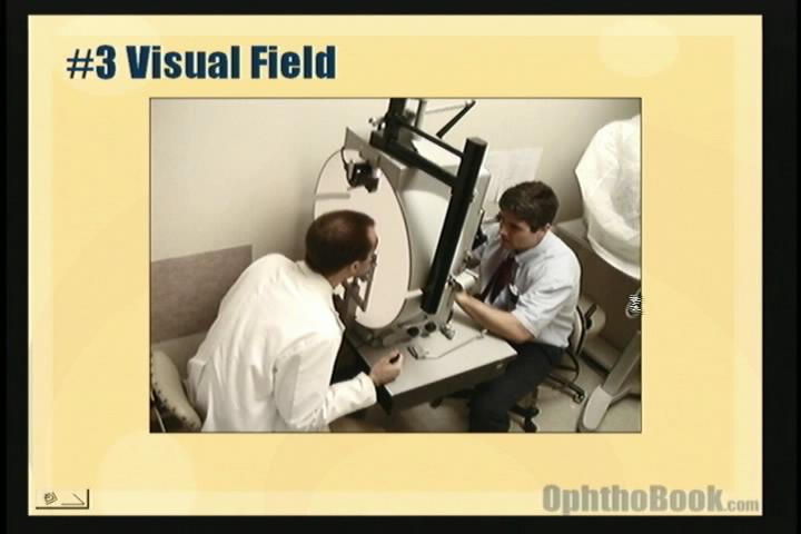
This Goldman perimeter lets us map out visual fields.
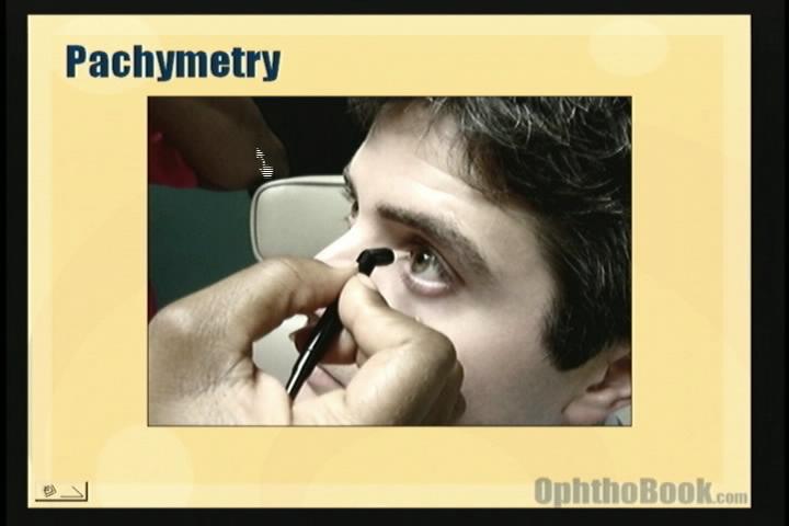
Pachymetry is the measurement of corneal thickness, and works through ultrasound.
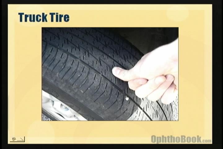
Some people have thick “truck-tire” corneas that feel hard at any pressure.
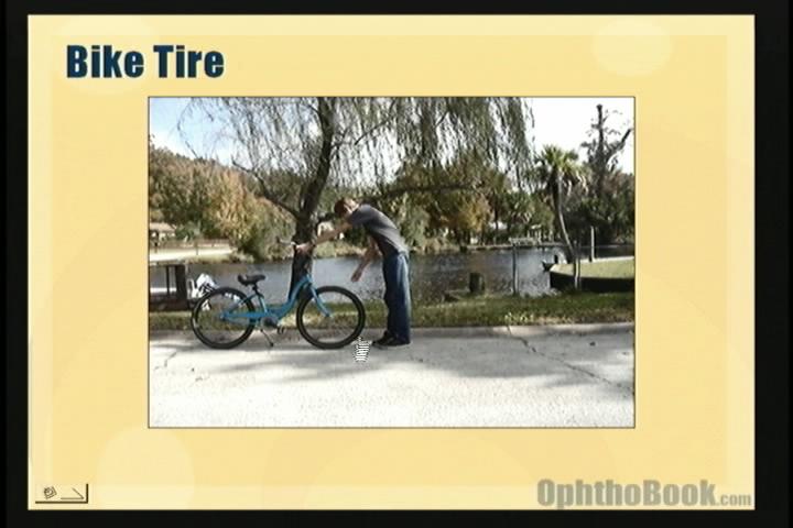
Other people have thin “bicycle-tire” corneas that feel soft.
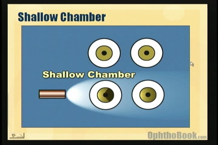
The chamber depth can estimated with a penlight.
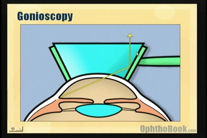
Total internal reflection limits our view of the angle and the trabecular meshwork. A goniolens breaks the cornea-air interface, and allows direct visualization of these structures.
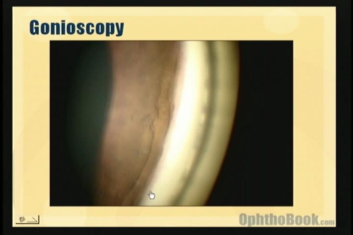
This gonio-video demonstrates the main structures and the trabecular meshwork can be seen as a faint line.
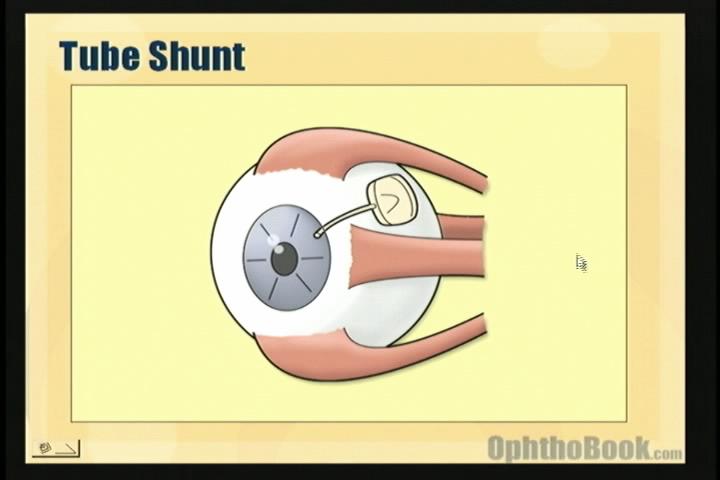
One surgical treatment for glaucoma is a tube-shunt drainage device.
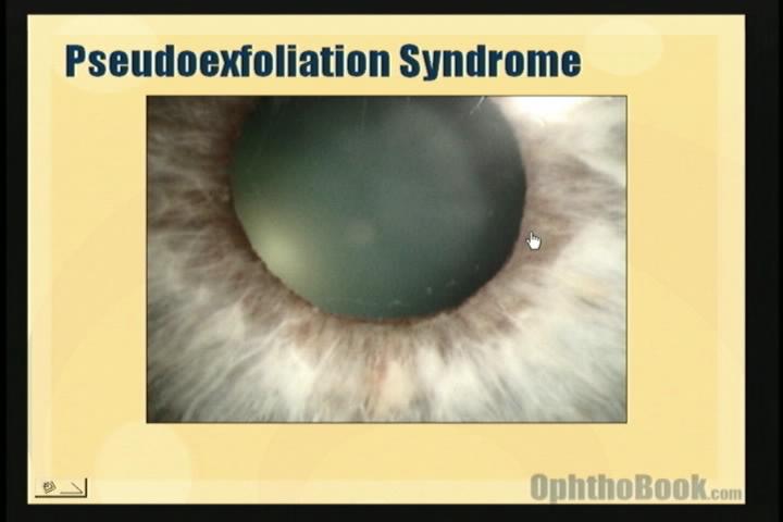
With pseudoexfoliation syndrome, a basement-membrane material forms on the anterior lens capsule. As the iris rubs against this material, pigment is scraped off that clogs the TM.
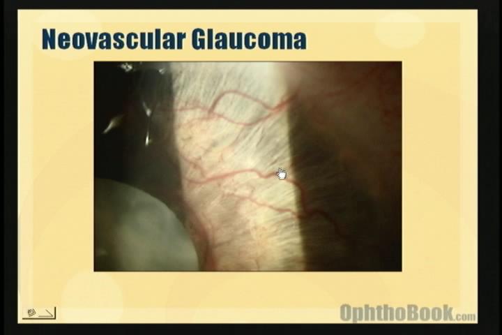
With bad diabetic retinopathy or a central retinal vein occlusion, VEGF can float forward into the anterior chamber and lead to neovascularization of the iris and angle, creating a dangerous glaucoma.
it is very usuful way of learning by waching this video.
i want video on different type of slit lamp technique ,needed for my exam.
Please tell me about the parts of the chamber when you take the gonioscope: Schwalbe line, trabecular meshwork, scleral spur,and the ciliary body . What is way differ bettween them to confirm the stage of close angle glocome
Hello Dr, congratulations for this website, it’s the most comprehensive resource in ophthalmology. It took me shorter time to understand many subjects than at the medical school!!!
gr8 job doctor..
very simple and efficient illustrations..the videos is so helpful ans handy..
i wish u the best in u’r life 🙂
love this video as i always like lectures with pictures demonstrating the things..student of MBBS .. i had to study the glaucoma from my boring and complicated notes but when i saw this video it became so clear to me that i just read my notes and i was understanding all the things so clearly….thank u so much…wish all the teachers explain things like u
waw 🙂 amazing video this what I need ..very useful
thanks a lot..
oh..no.. 🙁 why this video not work with me..
pleas can you send it to me at my emil..I really need it.
ok..now its work!I do not know why yesterday didn’t open with me!!!
thank you..
The videos are excellent, however the glaucoma one stops at 13 mins 32 secs for some reason. Great learning tool
wow!!!mind-blowing video.. very helpful
Hi, these videos are awesome. Thank you so much! For some reason the glaucoma video stops at 13 minutes 32 secs, as mentioned above…
this website is SO INCREDIBLY HELPFUL!! THANKS SO MUCH.
really nice nd highly informative……………
Incredible Videos….should be incorporated into all med schools curriculums
this is so helpful and thanks so much for your contribution to our education. i am sure we all benefit from this tremendously. thank you again for doing this!
Thank you doctor, i realy got the benefit…..thanks again
acan’t express my Greatfull towerd you
That’s an interesting video. Thanks alot, I’m a doctor. I teach and educate student in mylitary acedamy of medicine. I will use your video to teach my student
This is a very usefull lecture for students, residents and practicing doctors. Thank you very much for your time and dedication, you made it simple, understandable and practical!
Brilliant!!!
gr8 video!!!plz do add a note on d visual field changes in glaucoma..d rest of it.. jus tooo gud…
its superb!!!!!! thx doc
its realy very good explanation thank you but i wish if there is such videos for all diseases
what is blepheritis and what is the term for the inward growth of eye lashes
Glaucoma never made sense to me until today!
Hello, I really enjoyed watching your video about this topic and I would like to ask if you could send a copy of this to my email address since I would like to show this during my reporting on this topic.
It would really help me a lot to have such permission to have this video incorporated in my report. It is so comprehensive and I really like it a lot
Hi,
Thank you very much for such a wonderful video. Can you give me permission to incorporate this video in my library.
thanks for this good video….I understand many things easily
thank you very much
Outstanding 🙂 , THANKS A LOT !
Thanks a lot for this website!!! I’m an student of medicine from Spain, and I understand differnces between open and close glaucoma seen this wonderfull video.
It’s really comprehensive, and your english is really easy to understand.
Thx for your job.
WONDERFUL, EASY AND UNDERSTANDABLE LECTURE AS AN INTRODUCTION TO GLAUCOMA..
GOOD WORK..
This is great!!!! taught me more that lectures at medical school 😀
Thank you so very much. This is exactly what I needed to understand. I appreciate you going over the rudiments and helping me to grasp the basics and later expand. Thank you is not even adequate for the light bulb that finally went off for me. your explanations and visuals are not only interesting but easy to retain. I can be taught!
My favorite Ophthalmology e learning site on the internet. Comprehensive, subtle and yet brilliant! Thanks so much!
I love this site 🙂
Im gonna recommend this site and your book to everyone !!
Thank you so much.
thanks a lot
This is just amazingly perfect thank you and may God reward you with the best :))
I just saw this video once. but i can’t anymore. i have tried may be a hundred times refreshing one in the morning, afternoon and evening since 2 days but i cant see the video. then i figured out that the problem was with the website where you uploaded the video. (vimeo.com). Please i have a request if you can upload to youtube or any other video website before 29th of december because i have a presentation on glaucoma. and send me the link. thankyou a million times.
This is an excellent video. I can’t seem to sign up.
brilliant video!! thanx for sharing! 🙂
my god…awesome work by the doctorrs….an overview of glacoma has been captured….a million thanks to the contributors r thier work….
Please help !! The video is not working. ,!!
GOOD VIDEO THANKS
Simple, to the point, brief overview of a complex entity. Brilliant work!
very helpful
keep it up
you did a great job.
congratulations
Hi Tim-
Thanks for the wonderful videos and chapters!
You mentioned you had lasik? How myopic were you? I havent’ heard many ophthalmologists who get lasik on themselves.. i’m a -9.00 in both eyes- it seems my options are IOL or nothing?
would love to hear your opinion on this controversial topic!
Very nice. Very educational. And yes, entertaining lecture too 🙂
Thanks alot
Great job Dr.Timothy. Thanks a ton.
In the above lecture u mentioned that u had Lasik done. In case of Myopia the axial length is already more and in Lasik reshaping of Cornea is done by burning certain areas of it. I understand that due to certain pressure the axial length increases causing myopia. Does the chance of getting myopic power againg increases after Lasik? Please guide.
thnk u
i am not able to assess the video,can u help sir
thanks a lot sir
Thank you so much for website but the video is not workin.please help me
Thank you for your wonderful idea, I hope I have a teacher like you, the topic is well explained.Thanks again
this is an excellent stuff. but the problem is that none of the videos is running. how can I get them or watch them live
This is fantastic, i dont know how to thank you!
thanks Dr soo much this is a gr8 jop
i like it very much.good job sir.
glaucoma video is awesome too!! lovely lecture! you’re an awesome lecturer sir! thankeew.. =D
thank you very much with this video ! 2 months ago there was concluded that I have glucoma in 1 eye , only still can not understand why I can see 1 day only for 50 % procent through this eye and the next day is much better to 90% and nearly no black fields ! Any idea about this ?
amazing thanks alot
hello sir,im in luv with ur lectures.thanks a million for putting your time and effort to make these wonderfull, entertaining and very informative lectures.looking forward for many more lectures…..
god bless you for helping med students like me…
i just love this cartoonic vision videos make us v.easi to understand ophthalmology…
i just love this cartoonic vision videos make v.easi to understand ophthalmology…
Thank you so much .. I may love Ophthalmology becaous of you 🙂
I can’t watch the video frim An iPad…. Can you help me?? Thx!!
happy to learn dis way…nice
nice presentation.:)
Thank you so much! Finally someone explains things in a simple way 🙂
Video not play on my mobile I have Micromax canvas 2 help plz
very effective
Amazing! Thank you doctor
thanx,for great video
i cant thank you enough for making these videos. Im actually liking studying eye now.
Thanks alot doc!
please if you can email me this video. it seems to stop at the tonopen exam every time. love your work thanks a million
Thanks a lot! Excellent video, Easy to learn. Vevry-very useful.
wow .. I loved your biriliant way of explanation through the combination of pics & videos plus your simple comprehensive words .. simply you are outstanding Dr .. thank you
Thank you so much for all your wonderful lectures! I am a medical student in Nepal, so you truly are helping students worldwide. Really appreciate the time, effort and energy you put into your lectures.
a great and very useful video , thanks a lot .
keep going please !
I’ve spent the last couple of hours on your wonderful site watching video after video after video and had to leave a small note to thank you Dr. Root and anyone else who’s a part of this project. It is so helpful for me, even as a 3rd year OD student. I cannot stress enough how valuable all of your hard work is and how much I’ve benefited from it so far. I wish you all the best and hope that you continue this project because words cannot express fully how much I appreciate all of this.
All the best to you Dr. Root. May you be well and in good health always.
Thank you so much for these amazing videos!!!
Great job!! Thanx alot for all this
great work sir ,from KCMC TANZANIA ,enjoying very much.
thank you so very much.. wonderful explanation and fantastic images!
LOVE IT!!