Goldman Applanation Mires for Glaucoma (Video)
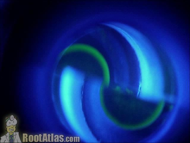
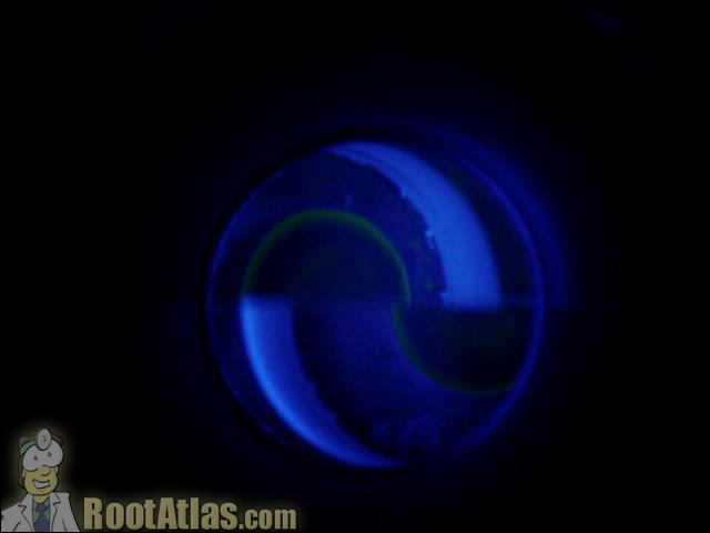
This video shows the view you have when checking eye pressure (glaucoma check) with the goldman applanation tonometer. This device flattens the corneal surface of the eye, and by determining the amount of force required to flatten a measured area, can calculate the inner ocular pressure.
Applanation is one of the harder steps for the beginning ophthalmology student, and can be tricky in patients with sensitive eyes and aggressive blink reflex. Also, it requires that the patient be able to sit up at the slit-lamp, which can be tricky in the infirm and the very young.
Goldman applanation tonometry is based upon the Imbert-Fick principle: such that the IOP measurement can be determined by the amount of force needed to flatten a fixed area of cornea. In this case, we are pushing on the eye with the blue-light applanator, and trying to flatten a round cornea surface with a diameter of 3.06 mm (that’s the area that our variable-force scale is callibrated for).
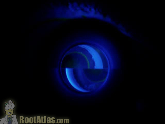
To ensure we have the correct sized area depressed, the plastic contact point has a prism in it designed such that you know you have the correct size area when the two hemispheres touch (like in these pictures).
Download this video
To download this video, right click on a link below and choose “Save Target As…”
applanationmires.wmv (2.9meg, Windows video file)
Screencaptures


I was surprised to find that the video of applanation mires is mixed up with anterior polar cataract. On downloading appalanation mires the video is that of anterior polar cataract! Please correct. Otherwise I am quite impressed with your site!
Dr Neelam Puthran
Editors Comments:
Sorry about that … I’ve fixed the download link. Thank you for pointing this out for me!
thanks, thank you, gracias. i can’t say it enough we now have a video resource to go to instead of odd description to blank faces for all our new staff.
tom
thanks , i was searching for the video of applanation tonometry for a ppt , Iam very happy to have found this site
Dr Rupa
Concise and easy to watch, thanks
It is a very nice photos and video but i would like to ask a question please
where should ,classically,i put the illuminating arm to the side of the eye to be measured or to the reverse or to the right side whatever which eye is measured or it does not matter?
it is very nice video,my doubt is fully clarified many thanks to you from my side keep on continue this help.
I have no idea who designed this website or took that photo but THAT IS NOT AN ACCURATE DESCRIPTION of what you should see when using the Goldman Applination Tonometer. The half circles pictured show the GREEN (as it should be) circles touching on the inner portion of each other and this is EXTREMELY INNACURATE. These half GREEN circles should just barley touch each other just on the outside of each other. If you were to use the IOP reading using this image you would have an inaccurate reading.
I apologize on behalf of the techs who do know how to measure the pressure. The video is accurate and well done.
No offense, Tech, but I think the picture and description on this page is actually pretty acurate. You want the INSIDE edges of the circles to barely touch each other for an accurate reading. That is what you see in this photo, and more importantly, what is demonstrated from the associated video.
The prism built into the applanation tip is set so that when a perfect 3mm diameter area of cornea is flattened, the INSIDE edges of the mires touch. It doesn’t make sense to lineup the outside edges of the mires … as the outside edge of those circles vary in size depending upon how much fluoresceine is in the tear film.
You may want to check to see if you have been checking your pressures correctly. Also, you might find it useful to run a google image search on the word “applanation” – you’ll see that everyone else is making the INSIDE edges line up as well.
Nice video specially for beginers.
Thanks
Regards
Shripat Dixit
It is one of the most useful instructive ophthalmolology videos. I am so thankful to you. Please keep it up.
Best regards,
Khalil Sheiban
More info please! Can you post a video of step by step instructions on how to use the Goldmann tonometer with dial control details from start to finish?! This would be extremely helpful for an assistant getting ready to test for the COA. Thanks in advance!
i just want to ask is the mire size accurate in this photo? the width of the mire should be like this?
Yep … width looks correct.
In general, the skinnier the mire width, the easier it is to line up and measure. This means, you should use as little fleuroscein as you can get away with. Put the drop in, have the patient dab as much out as they can, wait a little while, dab more … then applanate.
The hardest eyes to applanate are those filled with yellow dye and who blink a lot during pressure check. The dye gets all over the applanation tip and makes the mires too thick to read properly.
hi,
what causes the flouroscent semicircles to appear,are created by using the prism,how many prisms we use 1 or 2
Thank you sooo much for taking the time of making and posting the videos, they are SOOOO VERY HELPFUL..THanK YOU THANK YOU THANK YOU
Thanks much,you have clarified my doubts.
I enjoy your videos podcasts etc. great learning resources. Thank you.