RA – orphan posts

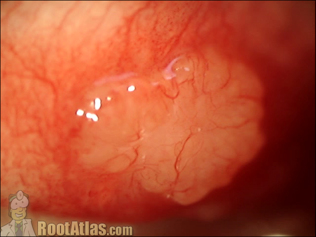
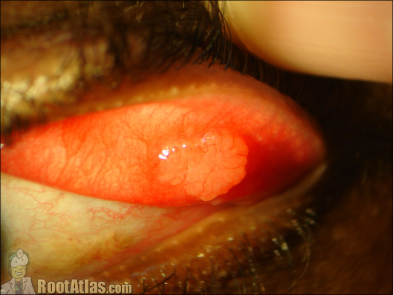
Photo: Sarcoidosis eyelid
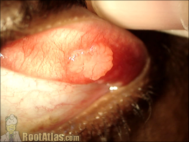
Photo: Sarcoid nodule on eyelid
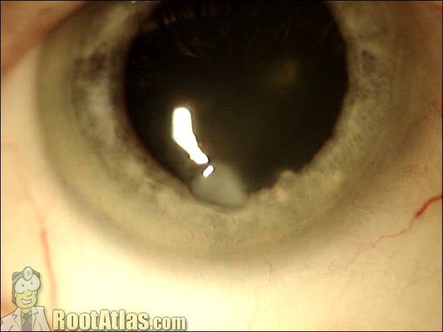
Photo: Salzman’s degeneration on the cornea
This photograph shows a corneal opacity called a Salzman nodule on the surface of the eye.
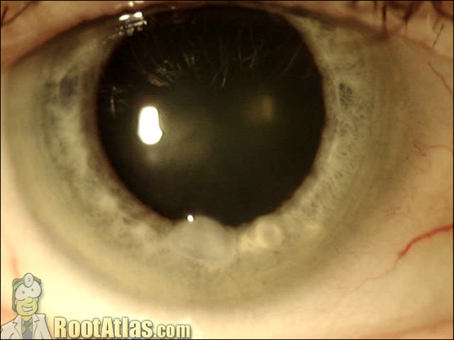
Photo: Salsman’s degeneration nodule on cornea
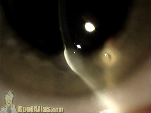
Photo: Salzman’s nodule on cornea
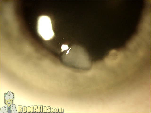
Photo: Salzman Nodule
Silicone oil in the anterior chamber (photo)
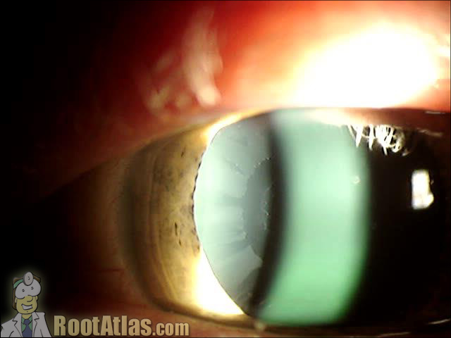
Pseudoexfoliation Syndrome (Photo)
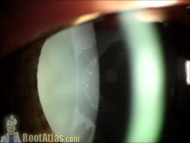
Pseudoexfoliation Syndrome of the Lens (Photo)
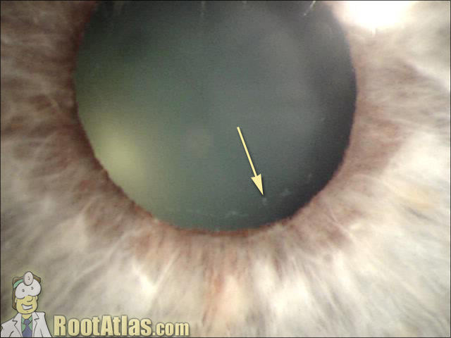
PXF of the lens (Photo)
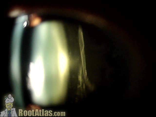
Posterior Vitreous Detachment by Slit-lamp (Photo)
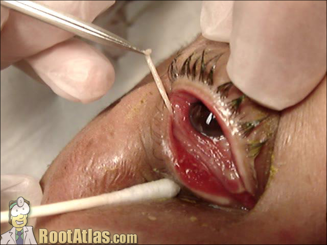
Pseudomembrane Removal (Photo)
e-eye.thumbnail.jpg” alt=”pseudomembrane-eye.jpg” />
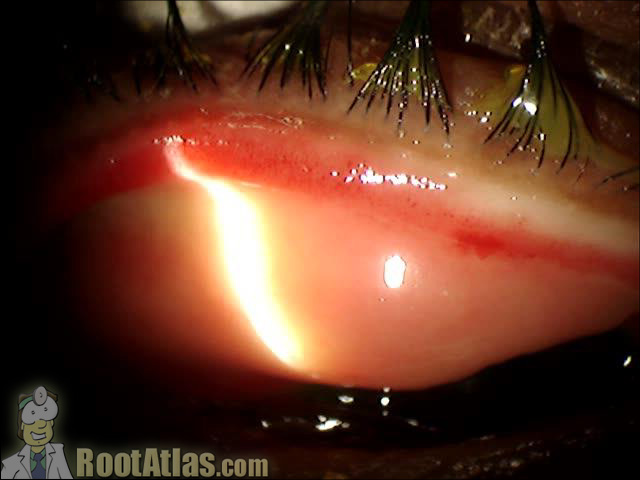
Pseudomembrane on Conjunctiva (Photo)
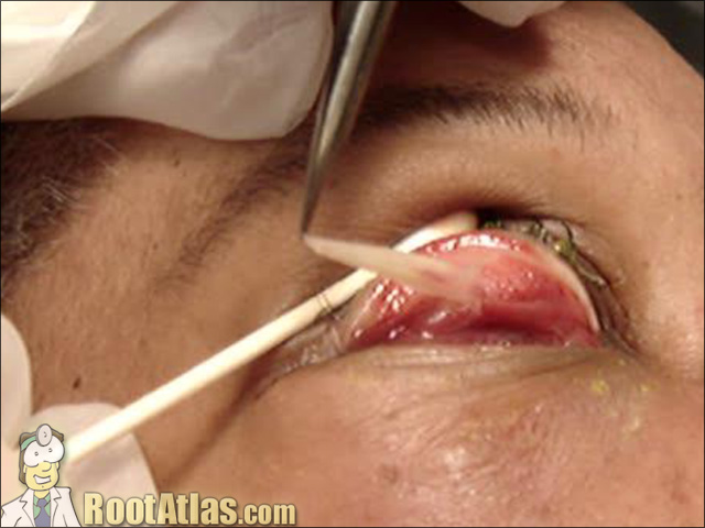
Pseudomembrane Removal (Photo)
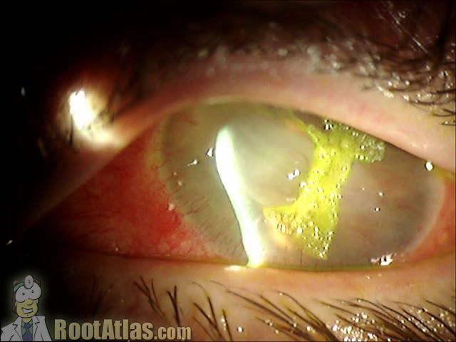
Cornea glue used to stop leak (Photo)
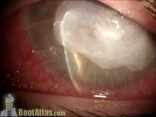
Perforated Corneal Ulcer (Photo)
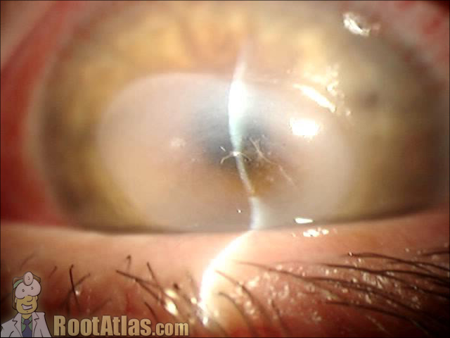
Desmetocele from Neurotropic Ulcer (Photo)
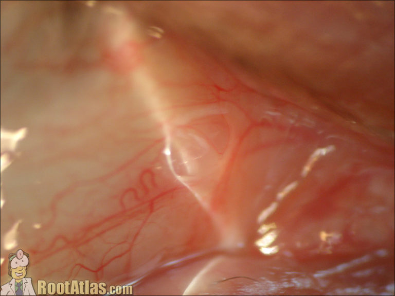
Small Conjunctival Cyst (Photo)
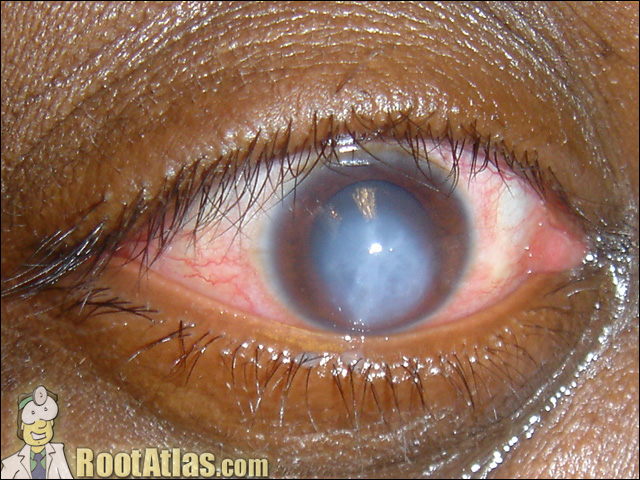
Keratoconus Hydrops of the Cornea (Photo)
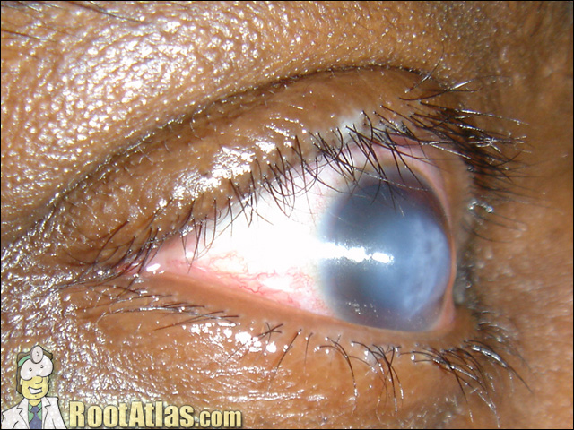
Hydrops Causing Corneal Edema (Photo)
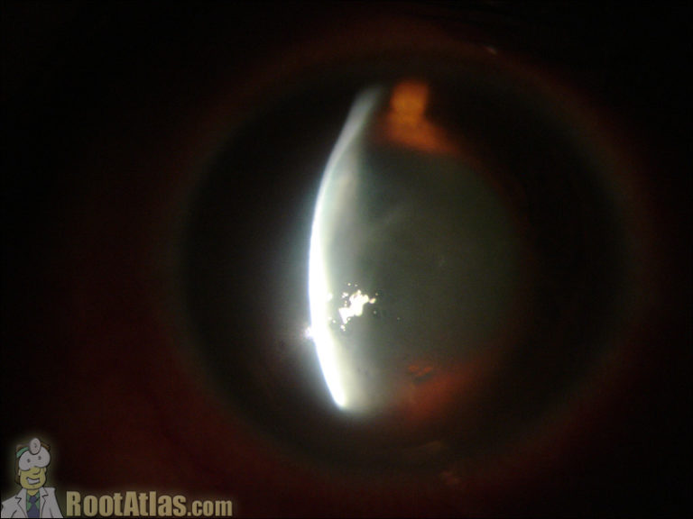
Corneal Edema from Hydrops (Photo)
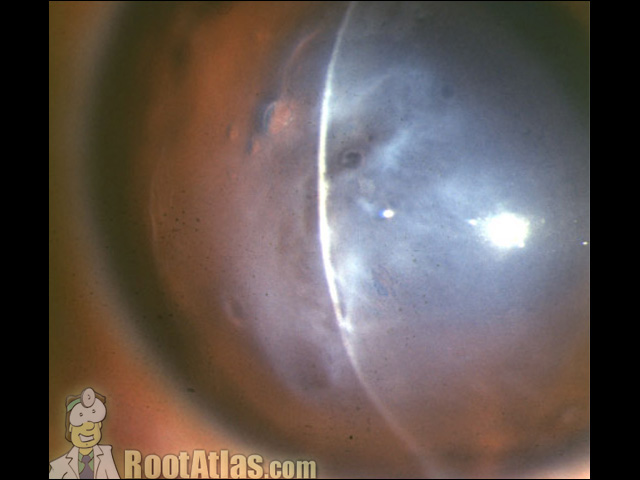
Hydrops from Keratoconus (Photo)
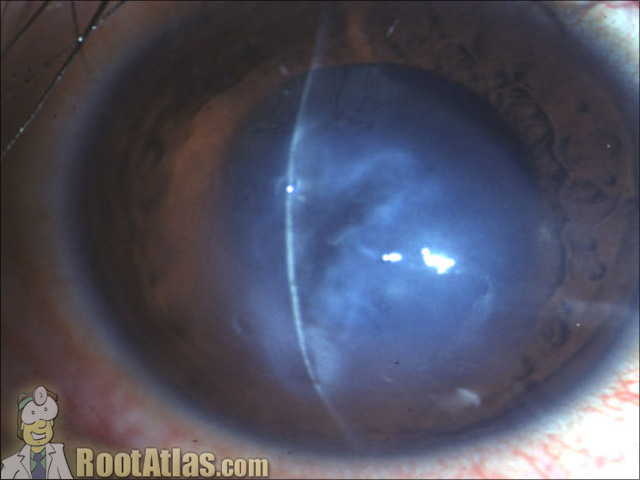
Hydrops Cornea Swelling (Photo)
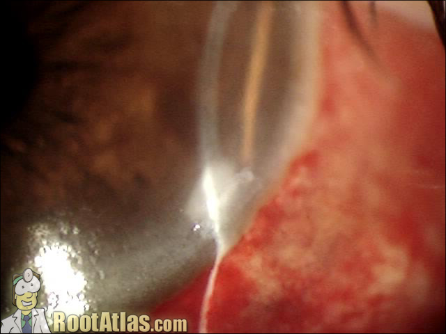
Dellen That Has Recovered (Photo)
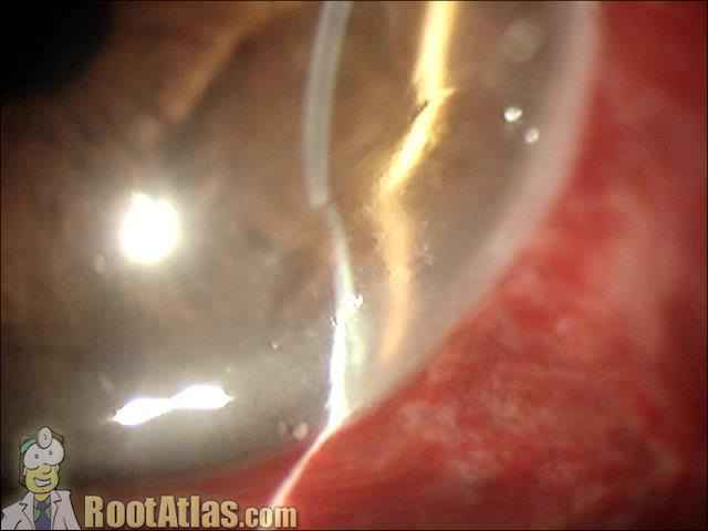
Dellen at Corneal Limbus (Photo)
This photo shows corneal thinning caused by a dellen (local dehydration of the cornea). Despite this “thinning” none of the cornea has been lost, it’s just dessicated. These occur from excessive localized drying. In this case, a large spontanoues conjunctival hemorrhage has formed under the conjunctiva. The conjunctiva is epecially elevated right at the limbus…
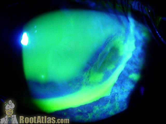
Corneal Thinning – Dellen Stain (Photo)
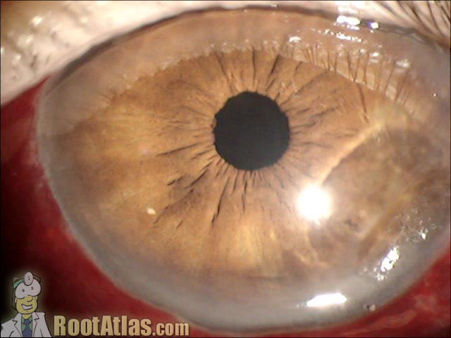
Dellen of the Cornea (Photo)
This photo shows a dellen (thinning of the cornea) that occurs from dehydration. You can see it at the limbus in the bottom-right of this picture. You can also see the funny reflection it causes on the iris underneath. Dellen occur when the tear film does not cover the eye. In this case because of…