Anatomy of the Eye Video
This second video is chock-full of high-yield anatomy facts. The eye is a complex structure with layers, lens, muscles, receptors, that is surrounded by many bones. I keep things simple in this video, and correlate directly with the anatomy chapter from the book. I’ve also scanned in an entire head CT to help you correlate the cartoons with real clinical imaging. Here are screen-captures from this video:
Screen Captures from this Video:
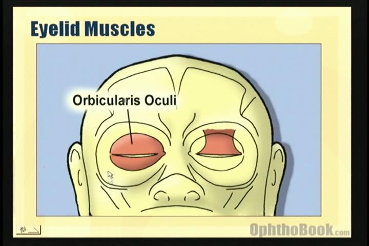
The orbicularis closes the eye, while the levator raises the lids. Each has their own innervation (cranial nerves 7 and 3)
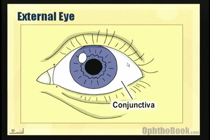
The external eye is covered by the thin conjunctival tissue, which inserts at the limbus of the eye.
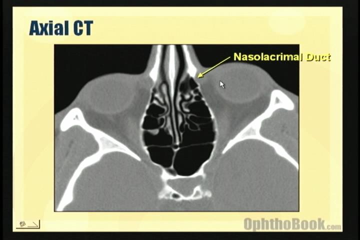
The nasolacrimal duct drains tears from the eye surface into the nose – explaining why your nose runs when you cry. You can see this duct on CT.
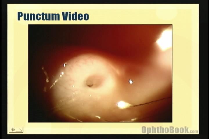
The punctum is small and located on the medial lid, near the nose. We can put plugs in the punctum to help with dry eye.
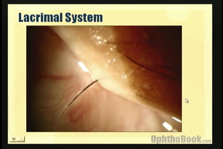
This patient has an eyelash that’s stuck in the punctum.
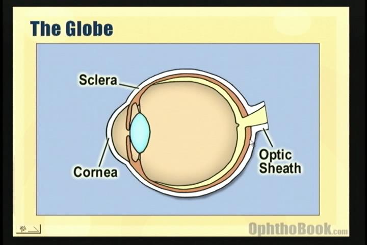
The cornea and sclera are continuous with each other … however, the cornea is clear because it is relatively dehydrated.
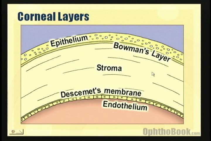
The cornea has five layers – the endothelial layer acts as a pump to keep the cornea dehydrated.
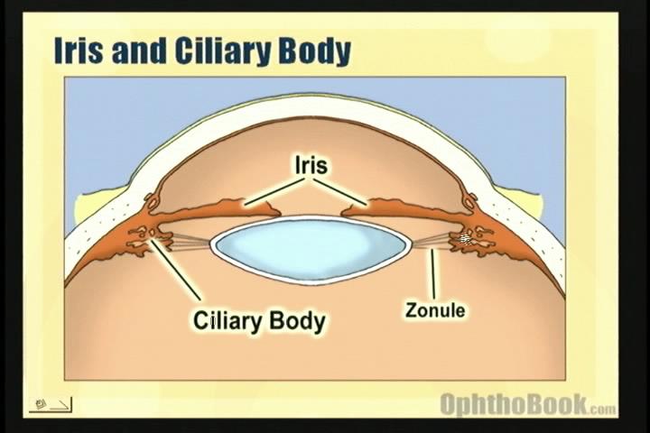
The ciliary body sits behind the iris and tethers the lens in place by a 360 degree network of zonular fibers.
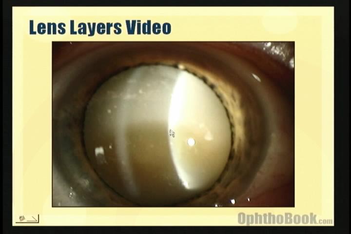
The lens has the configuration of a peanut M&M with an outer capsule, middle cortex, and central nucleus. In this advanced cataract, the cortex has liquefied into a milky consistency, and the central brown nucleus has sunk to the bottom.
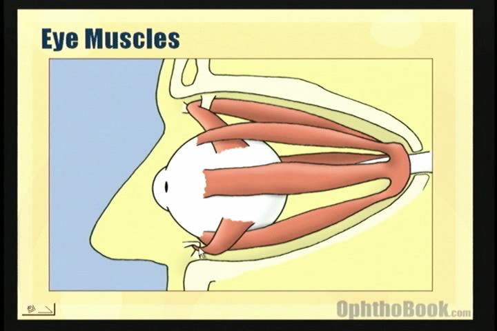
Eye movement is controlled by rectus and oblique muscles that tether the eye and connect at the orbital apex.
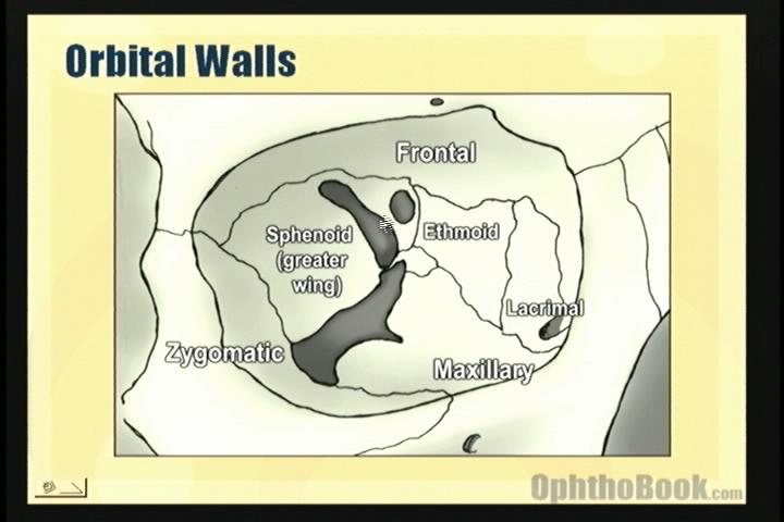
The orbital walls are formed by seven separate bones. They aren’t that difficult to learn when you review them one-by-one.
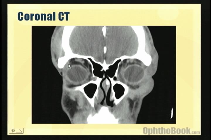
You can see the orbital bones and the extraocular muscles on CT – a coronal view like this one works best.
the films are perfect.
congratulations.
It is nicely explained.Good job!
its informative, good enough explainatory and very healthy education abt outer adnexa and anotomy in short time, excelent explaination ‘m looking more video abt this topic if u guys have just email me plz
thanx and best regards
this information is very use full students, very good diagrames simple points,
Thank you very much , this video helped me alot in understanding the anatomy of the eye … keep going ^-^
it is execellent i really enjoy iy
would like to see more!!
Thank you very much. This was great. It really helps students understand.
Superb! Thanks
i wanted to revise the eye anatomy before starting opthalmology 25 min worth watching the vidio thank you so much
this video was really very helpful….
hope everybody is doing fine.these videos are superb!but what should i do to download them?
I’ve been studying from books for my OMA certification but I’ve never been able to see the eye pictured like this. It has been a tremendous eye opening help (pun intended). Thank you so much.
Nice.
But where do i download this video ? is there any link to download it ?
It really helped me 2 understand the basics about the eye…pictures and illustrations were very helpful
I am a 4th year medical student. Currently attending eye ward @Hamdard University,Karachi, Pakistan. This video has helped me immensely and it made and impact on my memory (which is a very difficult job to do)…
I appreciate the simple examplainatory method and awesome slides. You have done great service for humanity…
Rasheed Syed 🙂
Awesome!a simple and methodical way.thanks for a quick revision.
This is one of the best medical websites I have ever seen.
the lecture was great and appropriately related all the clinicals.i think all students learning about eye should watch the video.
super ..when wil lens optics come..i’m waiting..
please put it up soon
Simply exellent.
Perfect video for ocular anatomy!
Awesome!
I am a student of B.Sc(HONS)Optometry and Orthoptics(ALLIED HEALTH SCIENCES) 2nd year in NISHTAR MEDICAL COLLEGE ,MULTAN, PAKISTAN.This video is especially helpful for the students of Optometry and Orthoptics.I am grateful to the site runners because.
Nishat Bokhary
Very good presentation. Thank you. This will assist me with my class lectures on Anatomy in a Surgical Technology class.
congratulations!!!!!!
I liked it, I Think that this kind of images are good like material.
One of the best things to learn anatomy is although pictures. that help me ….. thanks!!!!!
I am attending school for a Visual Impairment teaching certification, so I have no medical background. I am required to take an eye anatomy and visual functions class that has a very technical and confusing textbook. This video made turned gibberish and nonsense into something I could process! Keep it up and PLEASE make more videos!
Ilove the video and would like it very much if youl could send me the link through which i can download it so that it will assist me in learning my ocular anatoomy
The videos are a joy to watch and a welcome respite from the world of textbooks. Thank you.
fantastic lecture and great site
thousands of thanks from me to everybody connected to crate this…………thanks
MUY INTERESANTE, PERO SERIA MUCHO MAS BENEFICIOSO TENER LA PRESENTACION EN ESPAÑOL, SOMOS MUCHOS LOS OPTOMETRAS DE HABLA HISPANA QUE QUEREMOS APROVECHAR AL MAXIMO TAN IMPORTANTE MATERIAL… GRACIAS Y ESPERO SUS COMENTARIOS AL RESPECTO
thanks a lot. this video should be used as an introduction before hitting the core anatomy of the orbit and the eyeball and surely as a first chapter in an ophthalmology course. i hope you keep on doing this great job.
Thank you Dr. Root. brilliant and easy to follow video. Keep up the good work.
guys i wanna tell you that you are unbeievable-you give free amazing awesome lectures to all people just to make them understand-god bless you – my best wishes
azhar hussain
libya
Nicely Explained Lectures. Good job.
Hi, this is just great! =) Thank you very much.
thaaaaaanx………..i was confused of d interior
it is brilliant idea but ican’t acsses to see cartoon videos
Thank you so much.
These are all perfect,artistic.I have seen different videos from different resources,but watching yours,encourage me to study more.You know all what we need to know.
Again,this is ”art”.
it is great site for learning
Excelente, gracias por su tiempo para realizar estos videos de gran ayuda pedagogica
Hello,
That was really Great, I can’t express my thankfulness 🙂
Excellent job !
thank you very much for this excellent website.
Thanks for this.
Thanks for the video and website!!!
I would like to partner with you.
I am planning to establish an Online University for the poor communities of Madagascar. One of the subject I want to offer (not for making profit) is Ophthalmology.
Could you let me know how can it be possible?
Best regards,
Faly
thank u so much to all connected vid sharing this valuble knowledge n making optha more easy n interesting….
thanks recpectable persons
This is a very nice review course for boards! Thank you!
awesome lecture man
love it
thanks
xoxo
Really excellent work,I greatly appreciate your hard work and effort , I will give this site to my students to let them see how to be devoted to their work.
Thank you sooooooo much
what a video !
it’s amazing
go on…..thanks for your efforts
its very good and make everythings easy thank u very much
Excellent video;
Thanks a lot.
Regards from Madagascar
Great!
The cornea is not dehydrated relative to the sclera. It is actually the other way around, due to the cornea having ~4 times the number of glycosaminoglycans as the sclera. The hydration provides the proper spacing between collagen bundles to cause destructive interference, reducing scatter.
Great work thank you alot for the great efforts:)
thnx a lot ……….keep this work goin on…..it helps us…..
🙂 Well explained!
Excellent Video and great work , Cannot thank you enough Dr.Root
thanks a lot…m a medical student very useful…
Very good and helpful. Thanks a lot.
thanks a lot…
hey awesome video.
it cleared all my doubts.
can u please please explain development of eye.i find embryology really boring and difficult to understand.
thanks a lot
the video is clear for d understanding of d ug student
The video does not load on my computer. I am certain I have flash installed. Does anyone else have this issue too?
wonderful
thank YOU
I hope there will be more videos to watch with most common eye pathology.I am a general practioner and this concise videos are what I need and can change my practice thanx to you
thank you
very nice demonstration
loveeeddd it…your site is precious…
may god bless you for such noble services..:)
Wow, I am a medical student from Japan and I am enjoying these vids to the fullest. You are AWEEEEEEESOME!! I’ve already watched each vid x2 and am really really hoping you’d make many more of this!! Wish you could do this for all the medical specialties, you have so much talent, you are very funny, and keep the audience entertained. Never found optho so fun. Again, thanks for making such a generous contribution to all the world!!!! Will be sharing this site with others. LOVE
hi i am ophthamalogist in pakistan this video opened my fourteen closed doors. So please shared full knowledge of books and full subjects of ophthalmology. Thanks many times
I am indeed impressed by the great video and a very simple and comprehensive explanation. I am able to understand the anatomy of the human eye. Please more videos for other aspects of anatomy. Thanks.
very good video and helps to understand the sublect
I am indeed impressed by the great video and a very simple and comprehensive explanation.
Regards from intern from Palestine Al-Quds
thank you vetymuch
this is great
superb video easy way to understand ………………..how to download??????????????????????
It is a very useful site for medical students. Thanks very much for your contribution to us students’ learning.
amazing…thanks SO much!
great experience…wonderful site..thanx for being there..:)
im from bangladesh.
Great work, thanks alot for the great efforts:)
Pls how can i download your videos:i’ve watched the video on retinoscopy from a friends laptop,it was awesome.am a 5th year student of optometry in university of Benin Nigeria.
i m from somalia
i m medical student this is very usefull. thank you very much, you did great work.
It’s make easy to learn
im a medicine student from iran
many thanx for your useful efforts
Very good presentation
vary thanks for this video
I am a teacher of young children who are blind and low vision. I have been using your videos to upskill my colleagues on the anatomy and physiology of the eye so they can better understand the children’s eye conditions. Your style is clear and concise and that makes it accessible to everyone. Thanks and please do more! A series on common eye conditions would be very helpful!
this a great resorce to my projectcs im working on i might actually get an a plus
Thank you for great work
I hope u do lectures in instruments and investigations of ophtholmology
Exellent!!
WONDERFUL,VERY INFORMATIVE LECTURE, AND ALL THE ILLUSTRATIONS HELPED ME INDEED FOR BETTER UNDERSTANDING AND TO REMEMBER…THANK YOU VERY MUCH DR ROOT
My gratitude to Dr Root. I must say this is a wonderful resource ! So far one of the best i’ve come across !
hi there, i am from Recife in BRAZIL South America, and I really liked the sharing of this video with us down here in this part of the world…
thanks a ton !!!
It’s awesome! It was so boring to learn ophthalmology by faculty’s book. This one is much better! Can’t imagine my exam preparation without this stuff! Thank u, Tim! U r the best ophtolomology teacher ever!:)
I love it, you’re so cool
as a final yr medical student,i clear my concept of anatomy about eye,thanks a lot to give such awesome information
Thank you!!! From Trinidad and Tobago, West Indies
Amazing video! Is there a way to start the video at certain times? Clicking at a specific time just takes the video back to the beginning.
Thank you!
very great work
i am dr from somali i really intersting this lectures
it is very good learning method
Perfect and educative videos for med students and new residents ! Thank you for your willingness to teach !
Thanks so much for making this whole thing free. I’ve finish reading the book and would like to watch all the videos too. Unfortunately, almighty Great Wall of China don’t really like Vimeo, these video simply won’t load! Is there any other ways that I can assess them?
Agree with the amazingness, BUT like April 29th says, if you try to click on the timeline of the video it takes you right back to the beginning, which is VERY frustrating. If you want to rewind at all to hear something again, it’s impossible and will make you have to start from the beginning, and once you’re there, there’s no fast forwarding either.
thanks for this perfect video
Thank you for this. website… wonderful presentation…
I love this site! Having the visuals with the explanation really helps me understand what I’ve read about 100 times! Thanks for not charging for this info too!!
Excellent
THIS IS AN AMAZING CHANCE TO LEARN, IN DETAIL, THE THINGS I HAVE THE MOST TROUBLE WITH AS AN OPTOMETRIST’S ASSISTANT AND TECHNICIAN. THANK YOU SO MUCH FOR THIS WONDERFUL AND HELPFUL INFORMATION.
AWESOME WORK, MAN!!
Very well presented, quite Interesting well prepped!!
Great informative.
Nice….. and I Hope We need more n more from u…
Excellent! Thank you!
I absolutely love your teaching style! So fun and you make it so much easier to understand than the text books! I am currently studying for my certification and this helps thousands! Thank you so much!
Love your stuff! Everything is so great I supplement my new staff in off learning with your material. I have purchased countless copies of your book.
Awesome! Ophthalmology really made easy.