Retina Basics Video
This introduction to the retina covers all the basics: anatomy, diabetic retinopathy, retinal detachments, and macular degeneration. This is a lot of topics, so I’ve tried to keep things simple and to the point. Here are some screen-captures from this video:
Screen Captures from this Video:
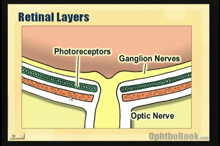
The video begins with a basic review of retinal anatomy. The key point here is that the photoreceptors lay pretty deep in the retina, with the ganglion nerve fiber layer superficial. The underlying choroid provides nourishment for the photoreceptors.
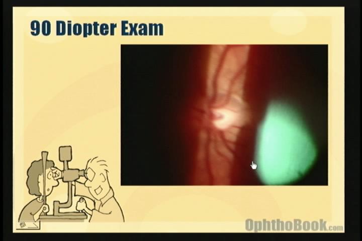
The slit-lamp is the best way to view the posterior pole. This full-motion video segment shows the kind of view you can expect when using a 90-diopter lens.
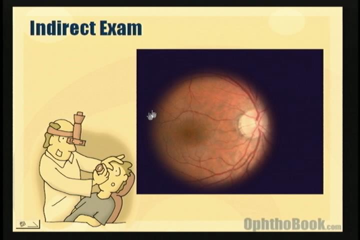
Indirect ophthalmoscopy is useful for viewing larger areas of the retina. The field of view is much greater and lets you look all the way out in the peripheral retina.
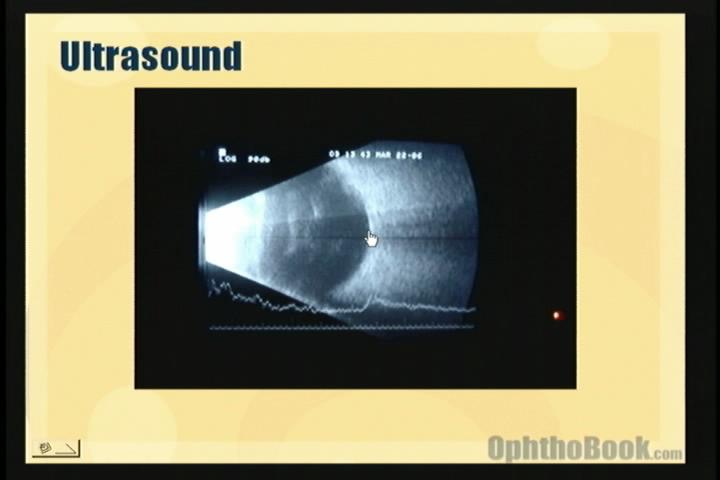
If you can’t view the retina (for example, the patient has a dense cataract) then you can visualize key structures with ultrasound. This full-motion ultrasound demonstrates all the important eye components.
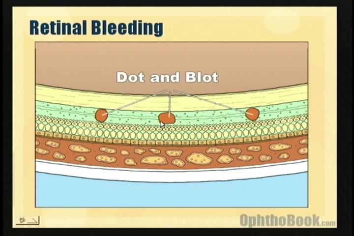
Diabetic retinopathy produces dot-blot hemorrhages. Dot-blot bleeding is discreet because they occur in the deeper, vertically-arrayed layers of the retina. Flame hemorrhages are larger because they occur in the superficial nerve-fiber layer.
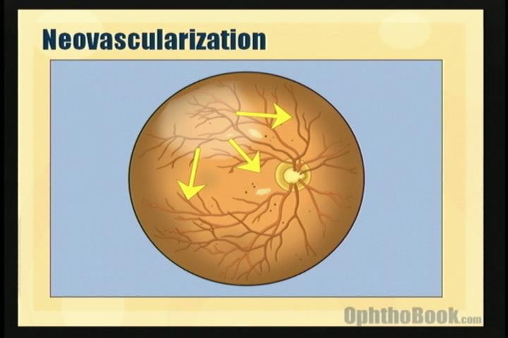
With large areas of ischemic retina, diabetics produce VEGF to stimulate angiogenisis.
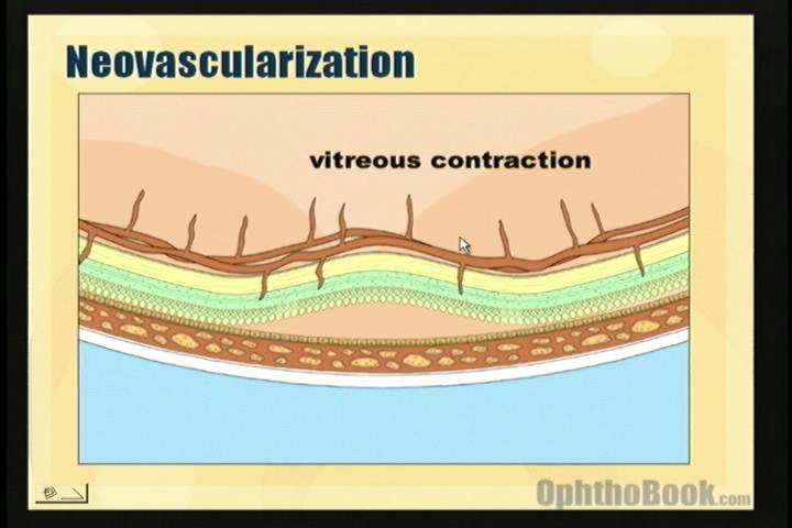
The new blood vessels can bleed and create traction.
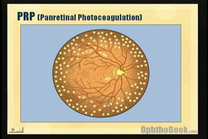
PRP, or panretinal photocoagulation is performed to kill off ischemic retina and decrease VEGF production.
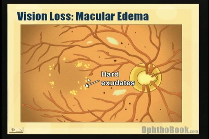
Despite other problems, it’s macular edema that actually causes of majority of diabetic vision loss.
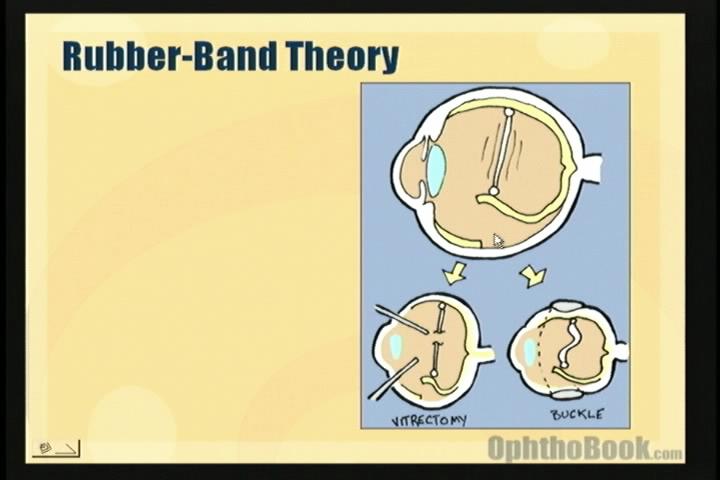
When treating retinal detachments we follow the rubber-band theory. You can relieve traction by cutting the band (vitrectomy) or by shortening the band (buckle).
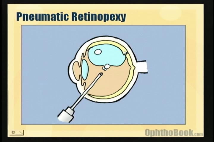
Pneumatic retinopexy can tamponade superior breaks.
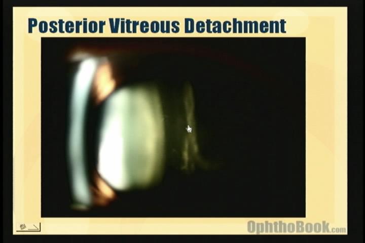
PVDs (posterior vitreous detachments) are common and can be seen at the slit-lamp in this video.
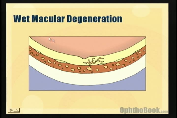
Macular degeneration is a leading cause of blindness in our country, and this video segment describes how it occurs.
It’s REALLY REALLY helpful , thank you very much 😀
good
hey, the retina video is not working (and all my friends at http://www.lf3.cuni.cz my faculty at charles uni 3rd fac i Prague have tried with no luck). So can you please post it again? or email me a copy n i can give it to the rest of my costudenst? please please please!! we all love your videos, it´s really helpfull.
Regards
Håvar Skoland
Editor: The video is working … I can view the video on every computer I have tried. However, this retina video is posted as an embeded windows video file, instead of flash video (the rest of the videos are in flash). If you’re having problems viewing this video, you may be on a mac system or have older hardware without the corect wmv codec support. Most PCs will automatically download the correct codec … but your institution may have blocked this functionality. I’ll eventually upload this video as flash as well, but this will take a long time and I’d rather work on the next lecture.
Thank you very very much, the page help me to do the paper for presentation. My interest topic is retinal detachment, it’s very useful and easy to read and understand.
Brilliant – Fair play to you!
Dear Editor: Macs are capable of playing the video you have inserted into this page, however, your HTML code is incomplete. In your code, you have “if !IE” – except that next tag should have been “embed” instead of “object” (object is for PC, embed is for Mac). Please feel free to email me and I can give you the exact code you can use to fix this problem. It is a 5-second fix!!! Thank you for all the work you have put into this website!
thanks so much doctor for the video..it is of great help..
but are there any videos that illustrate how to use the indirect ophthalmoscope using the 20 D lens?
video wont load !
Great video!
Simple and educational at the same time. Keep it up!
the video does not come up on the page
Yep i cant view it either… would love to see it. pls help! exam coming up 🙁
thanks for effort it`s so helpful
the retina video does not working
I’ve uploaded the retina video to Vimeo, so it should stream just like all the other videos on the site. Hopefully, you won’t have any more problems.
You may note that the sound is a little low on these earlier lectures. The later lectures are of higher recording quality (upgraded my system). Hopefully, the educational content will make up for everything.
hi
I just found your website (lucky me!)..Great video!it helps me a lot.I hope u’ll continue doing it.Btw, I do hope you reply my comment 🙂 thanks..
excellent presentation.thank you.
Thanks for watching??? More like thanks for uploading!!! Concise and precise. Can’t really ask for more! H
it’s really helpful.
thx.
Thank you, Dr. Root! You have no idea how many times your videos saved me from an endless spiral of confusion in my ophthalmology clerkship! Thank you again!
thank you Dr for this site and injoy by it is opthal learn with you
Thank you for this proffisional, didaktic and helpful media lecture.
thank u so much its very usefull
thank u so much… This video helps explain a lot of otherwise un-understood things.
Tank alot really helpfull website
thaaank you so much
u helped me alot in ophtha rotaion
u r the best ophthalmologist ever i hope if our teachers can tech us just like u
This site is amazing and so helpful. This is my go to source for my ophtha rotation and I am learning more from this than the actual rotation!! The notes are great, and the videos are even better!!
Thanks so much for taking the time to make this site and for making it free!
bored with long classess…. new way to get close to eye…..thanq very much
hey!! keep up the good work!! i have lots of doubts in ophthal..will it be possible for you to clear them for me please?
Top quality Videos, Thank you Dr Root.
Watching this in Optom School(final year)England,UK.
I have been using your videos to broadly review topics before and in between reading them up in full with your Pdf in addition to Kanski+ Taught lectures+ Tutorial notes.
Sugestion in making more Self Test questions/flashcards more accessible in pdf, please ???
Fantastic Work!!! Excellent!!!!
your videos are awesome. .its simple and easy to make out everything u teach. .i m an undergraduate frm india. .grateful
i treasure the moment i found this site…
really helpful…
thank you sir!
the retina video dont working please send for me
thank u very much these r vry nice and understandable
i found them vry vry useful want to download all
always simple and very enlightening
Thanks :)!!
generous professionals like you make our lives so much easier,thanks: a great summery of the important points clinically
Excellent review! Thanks soooo much!! 😀
Hello Tim,
I must confess that you are God sent! You are simply amazing and an expert in basic education.
Pls kindly post video of how to effectively use the BINOCULAR INDIRECT OPHTHALMOSCOPE. This technique is confusing to me.
Pls TIM help kindly as I enjoyed the tips on SLIM LAMP BIOMICROSCOPY!
Thanks in advance sir!
Thanks a lotttt, After a long search I finally got a best opthalmo teacher, My boards are comin n I found agrt book to study.Thanx a lot!!! 🙂
well this is a great video i am from
Recife – Pe, in Brazil south america and i think i loved this channel.
great work.
kudos to you man
seems like you should be in video making as well as being an ophthmologist.
The only prob being you can’t See it on an iPad or an iPhone
very interested presentation … we need more presentation on every topics in the ophthalmology to make it easy
So please can you arrange for lectures on Investigative Ophthalmology like Pentacm topography ,FFA ,US B-scan,OCT
etc…..
with regards ,,,, and so thanks for your great effort
Perfect!
If you can as well give us the video on schiotzs tonometry and visual field charting, we appreciate the most.
I watched every single video and they All seem perfect!!
I’m in my 5th year training in med. school and I have only 2 weeks of training for ophthalmology and these videos really helped me to take this training much easier ..
so THANK YOU VERY MUCH AND GOD BLESS YOU 🙂
help me to understand hypertensive retinopathy please.
Thank you for kind upload. Your knowledge about EYE is very good and help to understand more.
Thank you!! Simple, lucid and clear… as always.
Great video, easy to understand and informative. I’m a patient, not a doctor, and nobody seems to have the time to really answer questions and explain in detail. You have the video, pictures, and text to do that and much more. Now I can converse with my doctors in an intelligent way. Thank you!
well done. thank you
v informative.v nice
wowwwwwww
Excellent. Simple and educating.
It is a good start and thank you for the excellent work.Just wondering
If i could get some help to access slit lamp and 90 deg lens exam video.
I greatly appreciate your attention to my request.
Regards,
Rahim
you are the best!
excillent work,,thanx
Thanks Dr. Really helpful 😀
Thanks for these videos. Just bought the book.
You’re helping us oldies brush up on their revision also.
Thank you for your great videoooooooo 🙂
Thanks for these open source lectures! They’re incredibly helpful! I
Hey Dr. Tim, you’re a genius! I’m a medical student from India and found ophthalmology very very boring till now. But you goddamn owned it, fantastic teaching method and exceptional content. Thanks to you, ophthalmology is even more interesting than mainstream medicine now! Kusod to you and your effort! Looking forward to the Cataract lecture video.
Hey Dr. Tim, you’re a genius! I’m a medical student from India and found ophthalmology very very boring till now. But you goddamn owned it, fantastic teaching method and exceptional content. Thanks to you, ophthalmology is even more interesting than mainstream medicine now! Kudos to you and your effort! Looking forward to the Cataract lecture video.
Very informative and easy to understand. Thumbs up to you sir!
Excellent..keep up the good work!!
I’m having issues with visualizing the macula with the 90 diopter lens under the slit lamp. I can easily visualize the nerve, but I can’t get the macula. Do I move the scope nasally or temporally? If someone could answer this I would be appreciative.
AMAZING!!
Thank you from an MD4 in Australia!